
Short Communication | DOI: https://doi.org/10.31579/2641-0419/156
Department of Cardiovascular Medicine, Heart and Vascular Institute, Cleveland Clinic, Cleveland, OH 44195, USA
*Corresponding Author: amir R. Kapadia, MD Professor of Medicine Chair, Department of Cardiovascular Medicine Cleveland Clinic 9500 Euclid Avenue, J2-3 Cleveland, Ohio 44195, USA
Citation: Sohum S. Kapadia., Serge C. Harb., Samir R. Kapadia (2021) Is Treatment Inertia Anatomical Similarities Between Mitral and Tricuspid Valves in Sheep, Ovis aries, Hearts. J. Clinical Cardiology and Cardiovascular Interventions, 4(8); Doi:10.31579/2641-0419/156
Copyright: © 2021 Samir R. Kapadia, This is an open-access article distributed under the terms of the Creative Commons Attribution License, which permits unrestricted use, distribution, and reproduction in any medium, provided the original author and source are credited.
Received: 26 March 2021 | Accepted: 05 April 2021 | Published: 12 April 2021
Keywords: valvular heart disease; sub-valvular apparatus; mitral annulus; tricuspid annulus
With the recent rapid growth in transcatheter mitral and tricuspid valve interventions, it has become increasingly important to understand detailed anatomy of the valves. In this study, we investigated the similarities, differences, and associations between the dimensions of the atrioventricular valves in a sheep model, as sheep heart valves have a similar morphology to human valves. A systematic dissection of twenty-five sheep hearts was performed, with annular circumference measurement, and sub-valvular anatomy documentation. There was a significant association (r=0.865; p=0.007) between the circumference of the mitral and tricuspid annuli. Authors also identified significantly more chordae tendinea in the subvalvular mitral apparatus compared to the tricuspid valve (15.8±1.2 vs. 13.9±1.5; p<0.001). In conclusion, there is a significant association between the size of mitral and tricuspid valve annuli, and the morphology of leaflets and subvalvular apparatus is different between the two valves. These findings could have important implications in transcatheter device design, sizing, and optimal intervention timing.
Running Title: Mitral and Tricuspid Valve Anatomy
With the recent rise in percutaneous valvular interventions, it has become increasingly important to understand in detail the morphology and relationship of the mitral (MV) and tricuspid valves (TV) [1]. Tricuspid regurgitation is commonly present in patients with mitral valve disease [2]. However, there are no guidelines on the optimal timing for intervention in the setting of concomitant valvular disease, particularly when considering percutaneous therapy.
Although advanced cardiac imaging with echocardiography, cardiac computed tomography, and magnetic resonance imaging provide unique opportunities to study the valvular apparatus, imaging can sometimes be challenging and lack the spatial resolution for detailed anatomic definition [3]. Further, normal human valves are difficult to obtain for anatomical dissection. Thus, we used sheep hearts to characterize the MV and TV's anatomical details. Sheep valves are notorious for being anatomically similar to human heart valves. Also, sheep models are most commonly used in preclinical investigational device studies. Therefore, this study would provide useful data for the development of new transcatheter technologies [4].
Materials and Methods:
Twenty-five sheep hearts were obtained from Carolina Biological Supply (Burlington, NC). Sample size was estimated to account for variability in anatomy and size of the heart using previously published studies5. Hearts with any damage at the time of harvesting were excluded, and all dissections were performed by SSK and SRK. With the ventral side facing up, 1-2 inches was cut from the apex to identify the ventricles. A longitudinal cut was made in the left ventricle from the apex to the base between the anterior (APM) and posterior papillary muscles (PPM) on the free wall. The length and width of the anterior leaflet (AL), and lateral (P1), middle (P2), and medial (P3) scallops of the posterior leaflet were measured using digital calipers. The circumference of the annulus was measured using a string. The count and origin of the chords were visually described. The TV was cut from the acute margin between the APM and PPM of the right ventricle. The dissection planes and measurements were similar to those of MV. In addition, the septal leaflet (SL) and septal papillary muscle were included.
The measurements of the MV and TV are presented in Figure (1a). Although the circumferences of the MA and TA were not statistically different (paired t-test; p=0.11), the MA and the TA correlated significantly (r=0.865; p=0.007) (Figure (1b)).
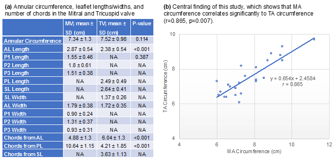
As shown in the Figure (1a), AL of the MV was longer and wider than the AL of the TV (p<0.001). Although P2 of the MV was largest in length and width compared to P1 and P3 of the MV, P2 of the MV was shorter and narrower compared to the PL of the TV (p<0.001). P2 of the MV and AL of the TV correlated significantly (r=0.607; p=0.001).
The posterior scallops P1, P2, and P3 combined had more chordae from the APM than the PPM. The AL of the TV also had more chordae compared to the PL or SL. However, overall, the MV had more chordae than the TV (15.8±1.2 vs. 13.9±1.5;p<0.001).
Within the tricuspid subvalvular apparatus, the APM did not provide chordae to the SL, and the PPM did not provide chordae to the AL. Within the mitral subvalvular apparatus, the total number of chordae to the APM and the PPM correlated significantly (r=0.703; p<0.001).
Very few studies have simultaneously assessed both atrioventricular valves within the same heart in terms of dimensions, geometry, and size correlations 6,7. However, these particular studies were solely based on surgeons’ observation in the operating room without quantitative analysis. Our study provides detailed in vitro measurements of each structure of the MV and TV, which can be used for future planning of transcatheter therapies. Although cardiac imaging is usually used to perform such anatomical comparison, it may not always provide an accurate depiction of the complex three-dimensional shape of the valvular apparatus. It also may not have the resolution needed to assess the subvalvular apparatus accurately8.
This article has some limitations. All sheep hearts were preserved in formalin solution. However, since we directly compared the MV and TV in the same sheep heart, the effects of formalin-related dehydration of the tissue probably affected both valves' measurements in a similar fashion without significantly impacting our findings.
In conclusion, there is a significant association between the size of the MV and TV annuli. The morphology of leaflets and subvalvular apparatus is different between these two valves, with more chordae tendinea in the MV. These findings could have important implications in transcatheter device design, sizing, and optimal intervention timing.
None
Dear Editorial Team, Clinical Medical Reviews and Reports. My experience with the journal was highly positive. The peer-review process was rigorous, constructive, and completed in a timely manner. The reviewers provided valuable comments that helped improve the quality and clarity of our manuscript. The editorial office was professional, responsive, and supportive throughout all stages of the publication process. Communication was clear and efficient, and any questions were addressed promptly. Overall, I found the journal to maintain high scientific standards and an excellent publication workflow. I would be pleased to consider submitting future work to this journal. Best wishes from, Elena Popa.

It was my pleasure to submit my testimonial concerning the Reviewer Board of our Scientific Journal “Brain and Neurological Disorders”. The Reviewers focused on some modifications and their contribution was helpful. The ladies of our Editorial Office were also supported my efforts. It was my honor to have such a co-operation and I am looking forward for more collaboration.

Dear Grace Pierce, Editorial Coordinator of Journal of Clinical Research and Reports, Thank you for the speedy and efficient peer review process. I appreciate the fact that your peer reviewers do not take months to respond like with some other journals. I would also like to thank the editorial office for responding quickly to my questions. It is an excellent journal. I plan to submit more manuscripts in the future. Best wishes from, Robert W. McGee

Dear Grace Pierce, Editorial Coordinator of Journal of Clinical Research and Reports, Working with you and your team on our recent publication in JCRR has been a truly wonderful and enjoyable experience. The responses were prompt, and the reviewers were patient, constructive, and highly professional. One reviewer in particular gave me the feeling that a professor was carefully reading and commenting on my coursework, which was deeply touching. The entire process was straightforward and hassle‑free, with no tedious online forms to complete. I highly recommend this journal. Best wishes from, DR Aibing Rao, Head of R&D

I Appreciate the Opportunity to Share my Experience with the Journal of Clinical Research and Reports. The peer review process was timely and constructive, and the feedback provided helped improve the quality of our manuscript. The editorial office was professional, responsive, and supportive throughout the process, ensuring smooth communication and efficient handling of the submission. Overall, it was a positive experience collaborating with your team.

Dear Mercy Grace, Editorial Coordinator of Obstetrics Gynecology and Reproductive Sciences, We would like to express our gratitude for your help at all stages of publishing and editing the article. The editors of the magazine answer all the necessary questions and help at every stage. We will definitely continue to cooperate and publish other works in the Obstetrics Gynecology and Reproductive Sciences! Best wishes from, Alla Konstantinovna Politova,
