
Research Article | DOI: https://doi.org/10.31579/2693-7247/084
1Kulvinder Kaur Centre for Human Reproduction721, G.T.B. Nagar, Jalandhar-144001Punjab, India
2Ex-Rotunda-A Centre for Human Reproduction, 672, Kalpak Garden,Perry Cross Road,
3Near Nawi Kachehri, Baradri, Ladowali road, JALANDHAR
*Corresponding Author: kulvinder Kaur, Centre for Human Reproduction721, G.T.B. Nagar, Jalandhar-144001Punjab, India.
Citation: Kulvinder K Kaur, G Alabamia, M Singh, (2022). ’An update on MRI of Brain of Neonates with its associated hurdles and Advancements for Analysis of Brain Morphology, Structural, Functional Connectivity and Functional Architecture for avoidance of Neuro developmental Diseases’’ -A Systematic Review’.J. Pharmaceutics and Pharmacology Research 5(7). DOI: 10.31579/2693-7247/084
Copyright: © 2022 Kulvinder Kochar Kaur1. This is an open access article distributed under the Creative Commons Attribution License, which permits unrestricted use, distribution, and reproduction in any medium, provided the original work is properly cited.
Received: 28 April 2022 | Accepted: 16 May 2022 | Published: 07 June 2022
Keywords: MRI; brain morphology; neuro developmental diseases
What is the method by which the generation of unbelievable cognitive capacity in addition to its working with the utilization of intellect.?What is the reason for5-10% adults for the generation of neurodevelopmental diseases or conditions like autism or dyslexia?For receipt of answers to these queries needs a greater insight in the context of the way human brain formation takes place in early stages . Studies with regards to models have not been adequate in view of variability of human species generation in contrast to other mammals in view of short time period of pregnancy in contrast to the duration amongst birth along with early adult period. Despite the brain of the newborn possesses an early along with comparative organization it being immature contributes to the postnatal modeling secondary to numerous experiences along with learning which the newborn infant’s exposure takes place. Furthermore the generating brain demonstrate a great plasticity subsequent to getting disrupted specifically with regards to the networks whose maturity takes place late at the time of childhood.
What is the method by which the generation of unbelievable cognitive capacity in addition to its working with the utilization of intellect.?What is the reason for5-10
Thus here we conducted a systematic review utilizing search engine pubmed,google scholar ;web of science ;embase; Cochrane review library utilizing the MeSH terms like anatomical; Quantitative(diffusion MRI), Multi parameteric strategies ; functional MRI ;In newborn ;preterm/fullterm infants from 1995till date in 2022.
We found 18,000 articles of which we selected 153 articles for his review. No meta-analysis was done.
Early generational modes
Prior to detailing the MRI techniques features significance exists to recall that longitudinal neuroimaging as well the measurements points to just an indirect reflection of the complicated steps of dynamic events visualized at the time of generation right through generation at the molecular, cellular, along with macroanatomic extent.The human brain generates at a slow pace varying from embryonic pregnancy to early adult period, dependent on numerous processes that take place amongst a remarkably restrained, however altering constantly [1]. At the time of pregnancy the brain illustrates a sequential generation of transient laminar chambers from the center towards the periphery;i) proliferative zones (ventricular as well as subventricular zones),the intermediate zone(later white matter(WM),the subplate,the cortical plate(latercortex )along with the marginal zone.
Neural proliferation, along with migration are common at the time of the 1st trimester of pregnancy, whereas,axon as well as dendrite growth take place basically in these cond as well as third trimester. Following that continuous maturation events are seen with synaptogenesis along with cropping modes, myelination, neurochemical maturation,etc.Allof these modes do not takes place independently however they crosstalk with maximum probability for prolonged time durations. Whereas early events take place endogenously directed by the genetic inheritance numerous of them were based on exogenous modes that have variation as per the baby’s milieu in utero along with subsequent to birth[2].
Macroscopically brain growth gets accelerated in the last trimester of pregnancy besides the first two postnatal yrs, with a considerable, escalation of gray matter (GM) along with WM volumes[3].The escalation of cortex takes place from -10to -150cm3 amongst 18 as well as39wks of gestational age(wGA;with equivalence to postmenstrualage (PMA) following birth )[4] along with from 200-600 cm3 amongst, 1 along with24mths of postnatal age[5].It is necessarily dependent on a logarithmical escalation of the cortical surface area;from --150cm3 at 27 w PMA to -700cm3as well as -2000cm3 amongst, 1 along with24mths of postnatal age respectively[6].This continues with the complicated nature of brain morphology in addition to the generation of gyri, primary secondary, along with tertiary sulci from 23,32 as well as 40wGA, respectively[7]. Despite the modes that are behind these get broadly debated[8].The folding of the cortex aids in the enhancement in the surface area with sustenance of adequate distances amongst the connections (as well as hence times for conveyance of knowledge amongst areas of brain.
These such potent macroscopic alterations in volume surface area as well as folding are possibly the marker that can get visualized as alterations in the microstructure of the cortical plate(latercortex), whose evolution in association with different modes at the time of preterm as well as post term durations. Subsequent to the migration of neurons the growth of communication amongst neurons to start with is vigorous as well as buoyant with synaptogenesis growth in addition to dendritic tree structure becoming complicated. Generation of the synapses is depending on fierce proliferation from 15 wGA,then on a phase of selective trimming of communications that are not brought into use that aids in just those communications possessing functional significance to get sustenance[14] .Simultaneously intracortical fibers acquire myelination,that is mostly at timeperiods of early postterm[15].These modes of communications, growth, generation of the synapses in addition to myelination take place over variable time periods based on cortical areas[1],with the primary unimodal as well as multi modal correlated areas that demonstrate variable paths of maturation.
In crosstalk with the generation of cortical areas structural connections get formed within the WM fierce along with interlaced events of growth as well as maturation at the time of pregnancy as well as early childhood [1,5,11]. At the time of the early preterm duration (26-30wGA),projection along with callosal fibers that possessed formation of communications amongst, subplate neurons initiate the invasion of the cortical plate as well as generate communications amongst neurons of the subsequent layer IV. Well generated limbic communications are existent in the cingulate, entorhinal as well as the hippocampal areas along with certain associative bundles (like inferior longitudinal fasciculus . At the time of the late preterm duration (31-36wGA),long along with commissural associative bundles get generated at a fast pace.During full term birth themajor long distance fibers existent have acquired their place whereas those fibers at shorter distance (likeU fibers ) get generated at the first year subsequent to birth[1].These anatomical communications get refined with certain reorganization via various modes that are complementary . Subsequent to abundant growth, communications that are not removed get deleted at the time of childhood via the cropping events as is seen for callosal fibers in non human primates[12].
Additionally events of stabilization of myelination the communications possessing functional significance escalation of the pace of transmission of messages amongst brain areas existent at a distance .In case of human brain this takes place from the 2nd part of pregnancy continuously till the end of adolescence, that peaks at the time of the first post natal year [15,4,13].The ages in addition to the rate of taking place is based on the areas along with networks with the common pattern of myelination depends on the caudorostral gradient, a propagation from the center of brain towards the periphery , in sensory as well as motor pathways prior to associative pathways.While the number of neurons along with microglia cells continue to be about stable in the post natal time duration the oligodendrocytes as well as astrocytes escalate remarkably in the WM. At the time of the first -3yrsattaining two thirds of the amounts estimated in adults[14].The hampering part of oligodendrocytes as well as myelin regarding neurite growth might partially reason out the lesser plasticity of the adult brain in contrast to the generating brain[15]. Noticeably, maturation occurs over various periods in addition to variation in rates amongst functional networks whose organization takes place earlier[16],with asynchronous propagation of myelination amongst brain areas [11,13,17].
Numerous neurodevelopment diseases get initiated from early interferences in these complicated as well as different modes at the time of the pre along with, perinatal time duration.This article in conjunction with neuroscientific work that target the insights of classical generation here as well as emphasizing the capacity of MRI for diagnosis besides prognosis of conditions associated with sulprematurity along with usual insults that newborn infant’s neonates incur.
General hurdles encountered with MRI of neonates
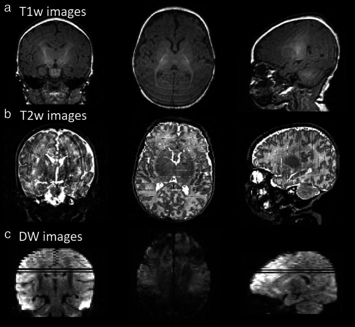
Utilization of 1.5 or 3’1’ MRI scanners is carried out in maximum studies where neonates along with infant’s MRI is implicated. Although greater significant correlated radiofrequency radiation ,3T neuroimaging does not result in induction of considerable escalation of temperature[19]. However numerous hindrances get encountered on dealing with this kind of patients population which renders research regarding brain that is generating possessing greater complexity.In maximum centers healthy full term infants it is not feasible to sedate with regards to research in mind however just with clinical reasons .This creates challenges as the images possess great proneness to movement(figure1[rev in 20]). Numerous strategies are utilized by various centers with certain groups making a choice of utilization of short acquiring sequences which can be run at the time of a protocol that is not greater than 30-45’ along with utilization of acquiring that possess tolerance towards movement in addition to strategies with regards to reconstruction that have got explanatory fashioned in this population where images are difficult to acquire[21]. Utilization of particular settings (like hearing getting protected with earphones specific for this or earmuffs ;that restrict the slope of the magnetic field gradient escalation )further result in reduction of scanner noise to certain limit besides provision of extra protection for ear.
Furthermore, the brain structures possessing smaller size creates problems along with escalation of the spatial resolution with regards to image is the requirement for avoidance of remarkableactions of partly voluming.Apart from maturation being not complete in the brain of infants results in separate tissue properties in contrast to adult brain causes variability in values of MRI properties(like relaxation time ; longitudinal relaxation time(T1) as well as transverse relaxation time(T2) diffusiveness gets estimated in diffusion MRI . Variability in homogeneities in signal as well as variation in these properties are seen across brain areas with the facts of asynchronous maturation as detailed earlier.This problem needs to get adjusted to the acquiring sequences for deriving germane in addition to enough contrast in the context of images . With the utilization of coils that are dedicated that posses ideal size as correlated with the head size that requires imaging, further escalates the feasibility of maximization of the signal-to- noise ratio(SNR) In the images [21].-with a greater than x2 , based on the brain areas along with their closeness to the coil elements .Taking into account the commited image post processing gadgets is the further requirement for tackling with the signal along with contrast specifications of the newborn images. Imaging studies of the brain that is generating thus has requirement of teams that have specifically received training apart from becoming expert with regards to both acquisition along with processing of the data[22].In the past 2 decades an escalation of clinical as well as research teams possessing attraction in this topic apart from formation of projects on large scale projects (likethe developing Human Connectome Project(dHCP)),that targets fetuses along with newborn amongst 20 as well as 44w PMA; known as the Utrecht Baby MRI Youth Project whose target group is amongst 30 as well as 44w PMA;,the Baby Connectome Project(BCP) whose target group is amongst birth along with age 5yr).This has aided the generation of particular techniques like studying aspect of infant movement[23].
With the escalation of articles in literature the aim of this review is documentation of more recent research as well as clinical studies performed with the utilization of variable methodologies with regards to neonates along with infants.
MRI Regarding Anatomical as well as Relaxometry
Generational Specificity along with Technological Hurdles
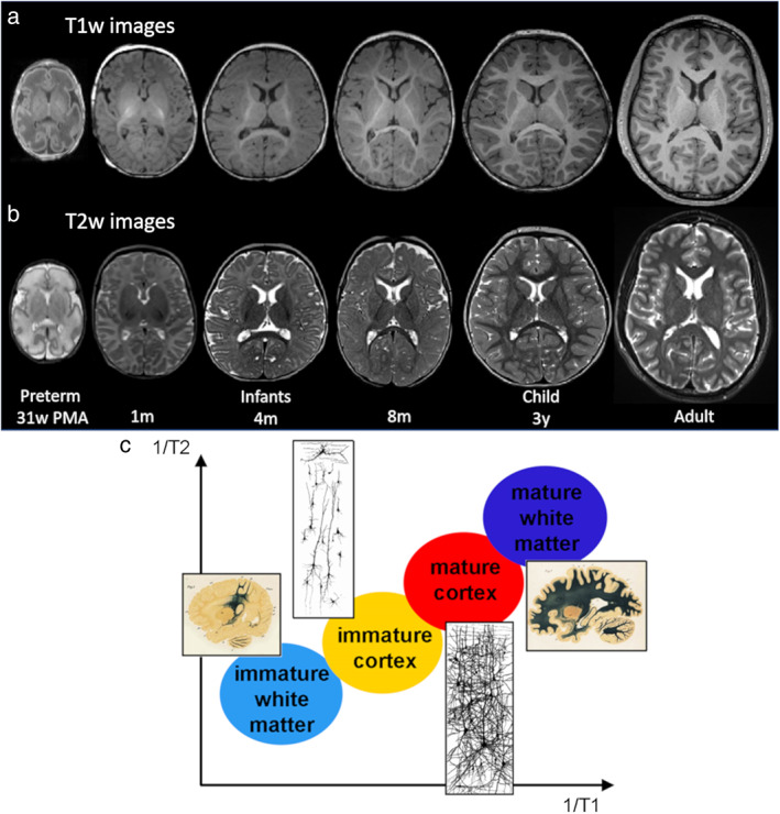
Regarding anatomical MRI that gets weighted by T1 as well as T2 relaxation time has to go via the hurdles of water along with fat amounts in the neonatal brain in contrast to adult brain that cause variable signal intensities In newborn along with infants.The evolution of these contrasts occurs with brain maturation in addition to further stages stepwise get usually detailed [11];i)theinfantile design(amongst 0-6mths) demonstrating a reverting of the normal adult contrasts (T1 weighted(T1w); lesser intensity of WM in contrast to intensity of GM; T2 w greater intensity of WM in contrast to intensity of GMii)the isointense design(amongst 8-12mths), possessing properties of a bad contrast amongst GM along with WMii)the early adult design(amongst >12mths) (T1w); greater intensity of WM in contrast to intensity of GM; T2 w lesser intensity of WM in contrast to intensity of GM(figure2a.b). Reduction of both T1 as well as T2 takes place with maturation events in conjunction with the reduction of water amounts.The alterations in T1 as well as T2 contrasts occur secondary to the reason that reduction of both T1 as well as T2 takes place with greater robustness in WM in contrast to GM in view of the myelination events(figure2c),to the degree that water molecules residing amongst the myelin sheath possess the least T1 as well as T2 properties [11].The time periods of reduction of both T1 as well as T2 are variable in addition to two separate modes might get differentiated in the WM the alterations in water molecules getting assorted (that influences mainly the reduction of T1 times at the time of the ‘’premyelinating ‘’state) along with the escalation of protein lipid with the chemical maturation of the myelin sheath that result in mainly in T2 shortening). .Hence the alterations are seen on T1 w images prior to T2w images.In case of neonates as well as infants at the time of initial 6-8 m post natal mths, T2w images usually get preference for outlining along with segmenting GM as well as immature WM,while utilization of T1 w images is for Identificationof the myelinated WM.The Objective of numerous studies has been to obtain ideal MR sequence parameters for enhancement of the image contrast amongst GM as well as WM.This involves specifically inversion times of 3D sequences at 3 T in neonates[24 ] as well as infants[25].
Furthermore, another hurdle for the correct outlining of brain structure is the resolution in space with regards to T1 as well as T2 images. Despitetaking into account 2D or 3D sequences, the provision of contrast is not essentially akin. With regards to research facilitation by certain groups the acquiring of images possessing great resolution2D images in 3 planes supportive for each other with slices that overlap(like axial,coronal,saggital )with the idea of utilization of so called ‘super- resolution ‘’ strategy in addition to re development of volume with greater resolution[26].The Objective of others is acquisition of images possessing isotropic resolution(-1mm) in 3 spatial directions [27].
Farther than acquiring data image processing in the context of anatomical images varies amongst premature newborn, infants as well as adult brains respectively. Different methodologies that have been devoted for this got posited recently for segmenting tissues, assessment, of morphology with regards to growth of brain , computating neuroanatomy of infant brain[28]. Combining images with variable contrasts( T1w as well as T2w) got evaluated for separatoin amongst tissues along with amongst, areas possessing variable maturation , however advantages are not clear cut thus far[29].Exploration of deep learning strategies got initiated however signal intensities across 1st postnatal year continues to remain a main hurdle[30].
For segmenting brain tissues, variable approaches posited whose classification is as i)unsupervisedii)parametric atlas fusion besides iv)deformable models[31]. With regards to the dHCP Project ,a totally automated pipeline has got posited in the context of processing images of preterm along with fullterm newborns from 28-45wPMA as well as provision of dependable extraction in addition to inflation of cortical surface[26]. Furthermore, longitudinal imaging can aid in tackling alterations in contrast in fullterm infants from 2w -18mth of postnatal age by integration of longitudinal hindrance along with provision of temporally constant as well as correct surfaces[28]. Greater volume as well assurface alterations in preterm infants from 30-40wPMAhave been seen in the occipital lobes in contrast to rest of lobes [32]. At the time of the initial postnatal mth, fullterm infants demonstrated, variable age correlated escalation of GM as well as WM volumes[29,33],that is for displaying the early fierce growth in GM in contrast to the greater prolonged growth in WM.
In conjunction with this the dynamic generation of cortical thickness in a spacewise heterogenous manner, however its estimation continues to be a hurdle with in mind the resolution of images in space.A commited pipeline of longitudinal data along with a multivariate assessment technique in advancement possessing the properties of temporal evolutionof cortical thickness from 1-24mths of postnatal age along with emphasized a generational regionalization in the context of structural as well as functional areas with meaning[34].Every one of these , illustrated a particular escalating , reducing pattern with thickness estimates varying amongst, 2 to 3.5mm along with peaks with thickness that is maximal with with variation in ages in the 2nd yr .By the time of 2years,mean thicknessis97% of adult estimates,while surface acquired is merely 2/3rd [6].Apparently possession of a little bit thicker cortices at 1 awa2 years of age (with a normal, range of estimates) might yield certain greater cognitive benefits in infancy as well as toddlerhood[35].
However the assessment of alterations in cortical thickness as estimated by MRI needs debate in view of them being dependent on images which undergo alteration as well as compared to maturation specifically at the time of the initial 2 postnatal years[3,11]. Initially it was pointed that the age correlated alterations in the seeming MRI thickness (with a prior escalation followed by reduction with age)might be correlated with over formation of synapses in addition to cropping . Nevertheless, synapses only project a little fraction with regards to the full cortical volume , along with modulation at dendrite,cells bodies, well as fibers levelmmmight represent the main parameter, impacting the estimated thickness[1].A combination of the estimated thickness with microstructural markers with regards to maturation of the cortex might aid in acquisition of greater insight of the events beneath. Moreover in view of the artifacts created by motion might differ with age well as bias,the seeming estimated thickness,quality of data in addition to quality regulation measures possessremarkable, influence on the age correlated directions [36].In toto the studies involving older children have pointed that seeming thickness on MRI undergoes reduction of maximum areas of the cortex by the age of 3 years [ref 37-rev in detail].
Brain folding Event-Mapping
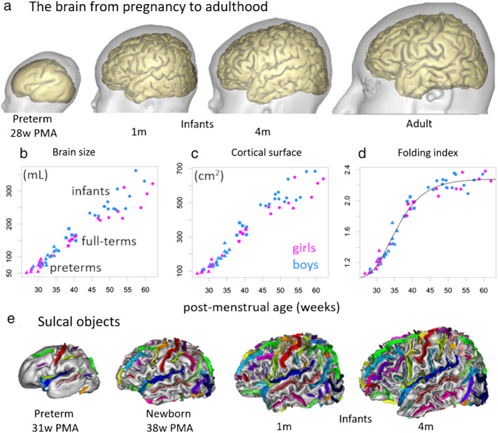
Depending on 3D redevelopment of surfaces of the cortex(figure3a),it becomes feasibleto compute abroad kinds of folding events(like depth,gyrification index,estimates depending on curvature)[27,38]. Evolution of these estimates occur in parallel with the age of the infant,via complicated modes with brain size as well as cortical surface area(figure3,b,c).In case of infants amongst27-62wPMA(-5mths of post term age) a nonlinear escalation of the gyrification index is visualized with main alterations at the time of preterm duration (prior to 40wPMA) with a reduction later[27] (figure3d). However,the gyrification index escalation persists subsequently at the time of theinitial auditory as well as visual cortices 2yrs , however at a lesser degree with greater growth areas in association cortices as well as lesser growth areas in sensorimotor[28].
Farther than the temporal issue studies validated heterogeneity with regards tos pace in folding across areas. The usual structure in space with regards to cortical motif got further described with a spectral assessment of gyrification with identification of 3 continuous waves across the 27--62wPMAduration,that might possess equivalence to primary, secondary, in addition to tertiary folding[27].This strategy might make it feasible for differentiating amongst, the folds that generated in a successive way on unique scale. Moreover ,maps in the context of cortical expansion have been emphasizing the variations in growth which are in agreement with the generation of new folds at the time of 27-38wPMA duration[39]. Amongst 30 wPMA along with postterm correspondent age(TEA)the main primary sulci demonstrated vigorous growth(figure3e) along with sulci generation earliest appears to be the maximum influenced by clinical factors like birth weight,multiple pregnancy or continued mechanical ventilation [40].
Like in case of studies in adults ,the utilization of techniques in newborns for quantification of growth of brain along with volumetry as well as morphometry essentially do not give outcomes that are confluent , that makes differentiation amongst, studies tough.Total corroboration of no techniques has been done in view of no ground reality along with since acquisition of MRI images in the neonates possess restrictions that are inherent with regards to contrast of tissues in addition to resolution in space. Hence biological assessment, of the outcomes need to get conducted with precaution.
Registration of Brain at Variable Generational Stages
Studies that have been brain depending on voxel dependent statistics(like for morphometry,fMRI) group assessment or group contrasts need conjunction of the brains of patients in a space that is shared by registration as well as normalization with space.This is specifically posing hurdles for brains possessing variable sizes along with folding designs . More recently certain strategies have got posited. Certain utilization of segmentation of tissue maps rather than raw T1w as well as T2w images in addition to, take into account these maps as well as cortical surface in the nonlinear event in the context of registration[41].Akin to that a benchmark dependent approaches aided in registration of the brains of preterms, newborns, as well as infants in addition to different databases of adults[42].The DISCO approach(diffeomorphic sulcal based cortical registration) utilization can get made for embedding hurdles in a utilization of registration scaffolding for starting the DARTEL step(diffeomorphic anatomical registration using exponential Lie algebra; implementation in SPM software [Matlab,Mathworks ,Natick MAJ)(figure6). Utilization of anatomically hindrance multimodal surface matching (MSM)further seemed to be dependable for provision of correct crosstalk amongst longitudinal cortical reconstruction of the infant’s that are same[31]. Utilization of these technologies can be made for setting up age- based templates along with spatiotemporal atlases, the way illustrated in 36-44wPMA newborns with MSM registration that gets guided by cortical folding[43].Finding the properties of anatomical differences in time based alterations in addition to disease associated changes needs equivalent greater resolution atlases that are commited to the generating brain[44].
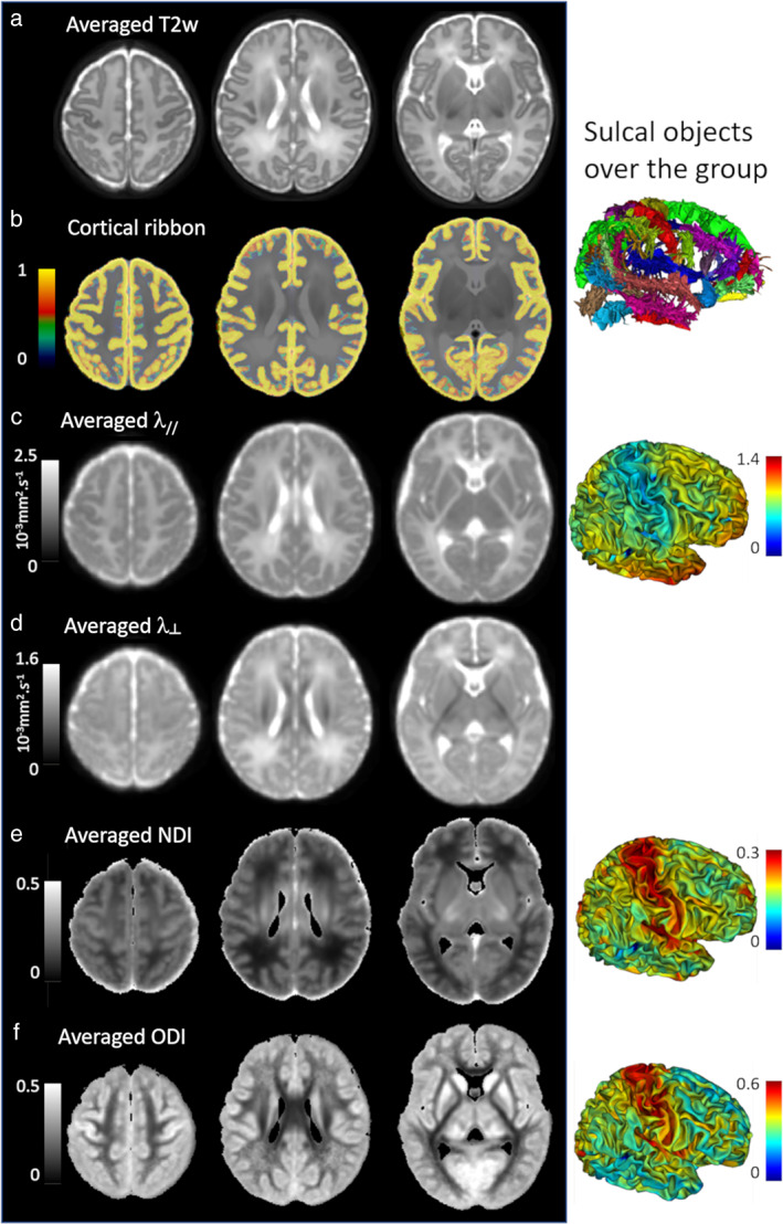
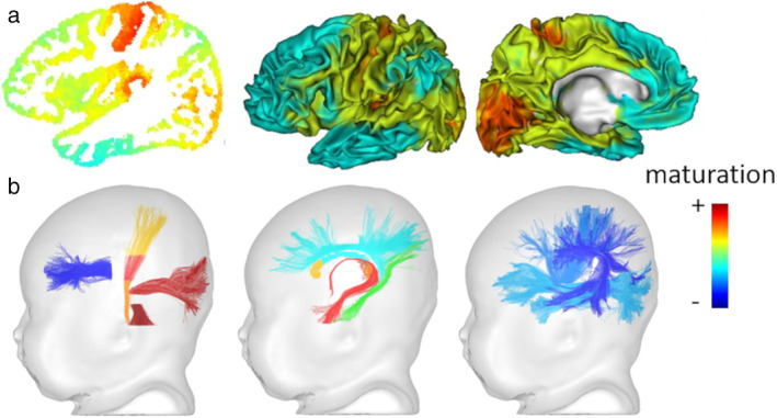
Courtesy ref no -19 Averaged maps over a newborns' group. The DISCO+DARTEL registration framework was used to register data of 40 full‐term newborns (first dHCP release: PMA between 37 and 44 weeks). The averages of registered images and maps are presented for T2w images (a), cortical ribbons (b), DTI maps (c,d: axial λ// and radial λ┴ diffusivities), and NODDI maps (e: neurite density index NDI, f: orientation dispersion index ODI). The right column shows the registered cortical sulci over the group as well as the averaged indices on the cortical surface, suggesting microstructural differences across cortical regions. 3.3D.Anticipation of the infants generation dependent on early Brain Morphometry
Various studies in the context of early growth of brain along with folding(at the time parallel to the third trimesters have been depending on preterms newborns,that cannot be believed to be a normal generational model.In variation of groups of preterm infants (like with or without brain damage ,intensely preterm -moderate preterm) in contrast to with full term infants[27,45]. Moreover,despite non consistence of outcomes, variation in growth of brain courses as well as morphology emphasis laid in fetuses as well as preterm infants without brain wounding [38]. However studies in preterm newborns is necessary from a Clinical point of view,as they might be possessing a diagnotic as well as prognostic part in babies who possess the risk of formation of sensorimotor or cognitive condition.Like Longitudinal brain morphometry in early preterm duration (from birth or at 30wPMA) as well as TEA has to certain degree aided in Identification of infants at jeopardy of cognitive along with motor dysfunction depending on machine learning strategies[46],despite socioeconomic status continues to be the maximum robust anticipator of outcomes .
Studies in the Context of Microstructure
In conjunction with morphometric along with morphological alterations, brain tissues illustrate main alterations in microstructural properties .Along with the generation of arborization of dendrites, myelination of fibers maturation alterations, in water amounts in addition to other events ,extensive T1 as well as T2 alterations, in GM along with WM[11,47]. However getting greater insight, with regards to, maturation events or interference with T1w as well as T2w signals can not get contrasted across areas or across persons in view of the variation amongst examination correlated with parameters associated with techniques like head size, along with position within the coil. For provision of this type of contrasts, either signals might get normalized for every patient,or T1 as well as T2 relaxation times might get quantification .
Initiatially it was suggested that T2w signals should get correlated with evey voxel towards the one of the local CSF that is not meant to differ amongst areas as well as right through generation[48]. Certain maturation was found amongst primary along with associative cortical areas correlated with language network in case of infants lesser than 4mths of age . In the recent past in the continuation of studies performed in adults,the T1w/T2w ratio has been pointed to a biomarker of myelination . In case of neonates this enhances the contrast of early myelinating WM structures (like posterior limb of the internal capsule , corticospinal tract,optic radiations)[49]. Amongst 36 as well as 44 wPMA the T1w/T2w ratio escalation further occurs in cortical areas that pointed to enormous maturation along with variation amongst, sensorimotor as well as associative areas[43]. quantification of estimates of T1 as well as T2 relaxation times represent greater righteous strategy for contrasting amongst, brain areas or amongst infants. Nevertheless for ths purpose acquisition of extra sequences that results in the prolongation of the protocol hence making it tougher for the undsedated infants. Various techniques have been pointed to more recently for the reduction of the time needed for acquisition. Utilization of 3D MP RAGE for( magnetization prepared 2rapid acquisition gradient echoes) sequence[50] or 3D SPGR(Spoiled gradient recalled) sequence with different flip angles[51], gives provision of dependable quantification, of T1 times. In the context of preterm newborns with imaging from birth to TEA, T1estimates illustrated a greater reduction with age in various areas of WM along with GM, inclusive of cortex in addition to central gray nuclei50,51]. Quantitative T2 times to be estimated gets aided byturbo/rapid spin echo sequences with multiple echoes ,whose reduction takes place persistently with escalation of age, in particular at the time of the first postnatal year (figure 2d)[52]. Spin echo sequences with various sequence parameter (various inversion timesor echo times(TE),computation of T1 awa T2 estimates over the total brain of infants in addition to greater resolution in space could get attained with the utilization of echo planar imaging(EPI)[53](figure4a,b). however the existence of EPI associated geometric deformation requirement of correction of the resultant maps in a nonlinear manner for the matching of anatomical images[54], along with provision of dependable estimates in the cortical envelop[55](figure4c).The utilization of BCP project alias a finger printing strategy for provision of concomitant quantification of T1 as well as T2 [56] . Considerable escalation ofR1(-1/T1) as well asR2((-1/T2)were found in WM areas from birth to -20 mths of age with subsequent slower enhancement following that.While assessment, in the context of maturation ,it is of significance to take into account their variation amongst areas in the mature state(variable brain areas might display variation amongst estimates in the adult brain).Just contrast of estimate amongst immature areas thus do not aid in coming to concluding correlated with advancements or postponement of maturation of one area in contrast to another [13].For performance of this, it is essential, to possess a reference for each approach estimated like on an adult cohort possessing a contrasting MRI protocol [53], or for estimation of their maturation courses as per age[55].

Specificity with regards to Generation and Hurdles in Methodology
In view of great water amount in addition to lesser myelination of the brain,the diffusion characteristics are remarkably variable(like greater diffusivity along with lesser anisotropy estimates(like variable in various directions) in case of immature brains of children along with , as well as adults[11]. Utilization of EPI sequences are usually made for acquisition of data along with recent advances of a multiband exaggeration method gives provision of greater reduction in the time needed for acquisition .While total brain diffuser tensor imaging(DTI) can get conducted in lesser time(<5>
Additionally,preprocessing constitutes a main step with regards to studies of neonates .Prior to assessment of diffusion results in a dependable method correction for artefacts with regards to movement is the prior requirement, if it involves intraslice or intravolume artefacts caused by sudden motion or floats diffusion seen amongst volume that are parallel to various gradient trajectories[60]. In the context of dHCP Project, a spontaneous processing pipeline got corroborated for tackling neonatal - particular situations in this large dataset[61].
Attempting the diffusion signal is further a challenge. With the knowledge of the needed acquisition time, despite that DTI remains the commonest strategies brought to use for the assessment of diffusion results of newborns[47](figure5a,b). however the correlated restrictions are multiple with Identification, of numerous deficiencies at the various stages of post processing[like seeref 62for detail review] in adults,that adds to be cautious in assessment, of the findings derived with this technique. Hence models that are more advanced have been suggested more recently for the assessment, of multiple shells HARDI results [47],like neurite orientation dispersion and density(NODDI)[63] (figure 6ef).or diffusion kuttosis Imaging (DKI)[64].Degradation of the signals into numerous constituents from numerous tissues with spherical denoising constitutes an appropriate strategy for provision of dependable longitudinal assessment of WM maturation [65].
Lastly as demonstrated for preterm newborns at 33,36 along with 39w PMA[66],with definition of age –particular diffusion template as well as atlases is the requirement, for consideration of special morphological alterations right through generation as well as conducted group contrasts or estimate aberrations in a dependable method .
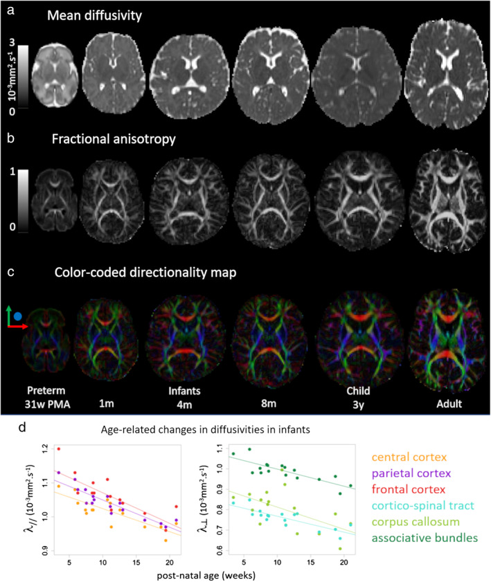
Diffusion MRI aids in a remarkable an accurate in vivo exploring of the GM. at the time of the preterm duration,the laminar organization of the cerebrum is feasible to outline as chambers like the cortical plate,sub plate as well as central gray nuclei demonstrated various diffusion characteristics of water molecules[51,67]. The evolution of these characteristics at the level of the cortical plate occur in a complicated method with age . Utilization of DTI, various groups have seen an early anisotropy along with a radial positioning of the major tensor eigenvector from 27w PMA, probably depending on the early existence of radial glia fibers along with apical dendrites of the pyramidal neurons [47,68]. Quantification of this temporsary microstructure got more recently conducted by a ‘’radiality index’’ with estimation of the local organization with regards to direction amongst the diffusion tensor along with cortical surface .Consequently, the diffusion amongst the cortical plate( achieves isotropy (like reduction of anisotropy )with elongation as well as complicated branches of neural communication (like basal dendrite of the pyramidal neurons,thalamocortical fibers).Thus the reduction in DTI anisotropy within the cortex apparently results in stabilization near the term comparable age while continuously reduction of diffusion indices is persistent [63](figure5d). Various microstructural modes that are competitive might offer a reasoning that is found in age associated alterations ,like reduction in neuronal density that is correlated with programmed cell demise , escalation of glial in addition to organelle cells, neuropile (alias a dense network of neurons and glia in the central nervous system). amongst cellular bodies getting complicated, reduction in water amounts etc instead of over generation along with cropping of synapses which are possessing relevance to just a little volume amongst the GM.The utilization of NODDI model get done for acquisition of greater insight in the modes at the time of formation [63],in view of the neurite density index provides information in the context of cellular along with organelle density in addition to the orientation dispersion index with the geometrical microstructure . Furthermore, provision of key understanding with regards to cortical microstructure is done by DKI withcontinued reduction in average kurtosis across the preterm duration[68]. Greater requirements of studies is existent for systematically contrasting the provision of biomarkers by the various diffusion models in the generating brain as per the age of the newborns in addition to the cortical areas.
With the utilization of studies having practically convergence of complicated models for demonstration of variations in microstructural alterations amongst cortical areas across the preterm duration[69], with a seemingly lesser complicated microstructure(ie greater anisotropy with regards to DTI along with diffusivity estimates) however images possessing greater intensity in the context of age associated alterations in gyri in contrast to sulci , in addition to frontal lobes in contrast to occipital lobes [47]. Moreover, the occipital lobes illustrated the greater maximum rapid alterations in the ’radiality index’’ [69] as well as kurtosis(alias the degree of peakedness or flatness of a probability distribution, relative to the normal distribution with the same variance [89]. Subsequent to 38w PMA, the enhancement of NODDI neurite density index is further seen , however is just limited areas(primary motor along with sensory areas )[63].
Utilization of DTI along with NODDI models the existence of heterogeneities in cortical microstructure have been found amongst the cortical areas of the total brain in full term newborns(figure6). Besides that certain variations have been detailed amongst the functional system like auditory along with linguistic areas of preterm newborns amongst 26 as well as 42wksPMA[70] along with amongst 1 as well as 5mths age[71]. Contrasting amongst the microstructure of akin cortical areas over the left along with right hemispheres that had voxel wise dependence have been made feasible in infants right via a cautious registration of local along with inverted brains as well as DTI maps with a 2 step matching approach of sulci in addition to cortical ribbons with the idea of compensation with regards to morphological asymmetries[72].This scaffolding emphasizing that asymmetrical microstructural organization in particular in sensorimotor along with language areas .
Despite, associations have not been demonstrated till now the complicated alterations in cortical microstructure the way evaluated with DTI along with NODDI might be possessing association with the gyrification events [63] along with maturation of the correlated WM tracts[68].These observations apparently vary amongst preterm infants at TEA along with full term newborns[70]. DTI further aids in the structural survey of central GM in the generating brain [47,51,73], however even now provisions of few evaluations with greater exaggerated models like NODDI or DTI are existent
Apart from the study of GM microstructure, diffusion MRI is the method preferred for mapping the generation of WM(connectivity along with maturation) in newborns as well as infants [11].
i)Connectivity of white matter
Even early in the preterm duration[74]the clearcut outlining of the major bundles, besides its organization on DTI maps with regards to direction, that demonstrate the major direction of the diffusion tensor (figure5c). More recently HARDI models along with determination of fiber orientation distribution function(fODF) have aided in the precise watching of fibers that cross like in corona radiate [75]. Utilization of broad categories of tractography techniques can be feasible with the idea of reconstruction of the seeming course of the bundles of WM in 3D. Nevertheless, those dependent on simple diffusion models (like DTI that just takes into account one single fiber population/voxel)pose numerous restriction that result in biases like false negative(preterm ending of a tract)or false positive(like shifting of a tract to the adjacent ones )[76].Just by the utilization of devices possessing greater advancements besides in need of HARDI data with fODF determination, by which hopefully structural connectivity investigations is feasible with the maximum dependence anatomically .However greater acquisition times are the requirements that in certain times is tough to attain in the context of newborns .
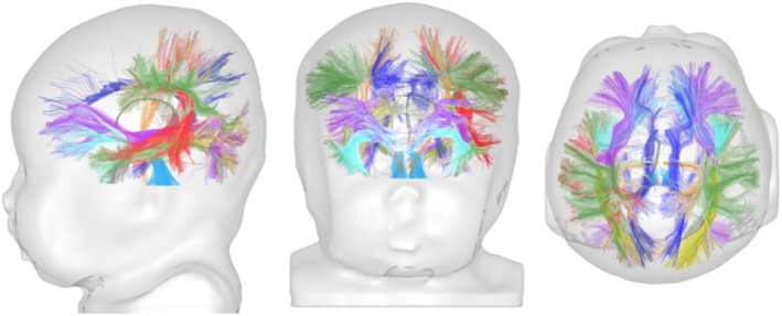
In case of newborns along with infants, present technologies, have aided in reconstruction of major bundles inspite of them being immature [53,77],the commissural fibers of the corpus callosum limbic bundles(fornix besides cingulum ),projection bundles(like corticospinal in addition to spinothalamic as well as optic radiations , anterior arm of internal capsule ) as well as associative bundles (exrternal capsule, uncinate fasiculus arcuate,inferior as well as superior longitudinal fasiculus)(figure7). At the time of infancy as well as toddlerhood, morphology of the bundles continues to be stable with a population particular utilization of atlas is feasible with the purpose of Identification, with Diffusion MRI [78]. Automated tractography kits that are inclusive of like earlier knowledge with regards to anatomical adjacent pathways,have further got in particularfashioned keeping newborns in mind [77]. However the biological assessment of tractography reconstruction continues to be a problem ,as besides the participation of axonal fibers, radial glial fibers as well as blood vessels also participate specifically in the preterm duration[79]. Additionally tractography technologies dependent on diffusion MRI do not aid in the estimation of cropping of axons[75],that continue still the intial some mths subsequent to birth[1].This event could result in alterations of signals, however these might not be apparent, in view of other alterations in opposite direction basically correlated with myelination might be the dominant factor. In future making utilization of benefit of the differential rates of maturation of bundles at the time of infancy for assessment, of their course with more dependence in addition to specificity [75].
More recently,the generating architecture of structural networks has further been described by total brain connectome strategies[80],which depend on connectivity matrices estimating the level of communications amongst, brain areas whose existence is in pairs.From 30wks PMA,the presentation by structural connectome is a ‘’limited world’’ modulation of organization like in adult brain [81] as well as certain cortical nodes (alias the ‘’hubs’’) are extensively communicated along with generate an ‘’affluent club’’ architecture [82]. The refining of this topology(alias.the way in which constituent parts are interrelated or arranged) are further achieved with age[81] possessing an enhancement in total effectiveness, along with incorporation as well as separation along with a considerable hieachial order right from primary towards greater order areas. However these connectome methodology in addition to methods of tractography reconstruction possess probable biases which alter the biological, correctness of findings the way described in adult brain[83]. Specific studies dependent on DTI possibly detail scanty networks in contrast to true ones as detailed in animals that could result in avoidance, of the correct outlining of ’affluent club’’ architecture in newborns[84].
ii)Maturation of white matter
Once the formation of connections occur the WM fibers with time acquire maturation in addition to function via the event of myelination which stepwise can get estimated with the utilization of Diffusion MRI [11,47]. At the time of preterm duration DTI diffusivities reduction occurs whereas escalation of anisotropy takes place in majority of WM areas[67,69].These DTI factors keep on demonstrating potent alterations in the initial post natal mths in Identification of bundles by tractography [53,85] (figure 5d). They probably possess sensitivity to various modes,like the proliferation of glial cells ,the continuation of oligodendrocyte projections along with them winding the axonal projections . Models of maturation with regards to findings in fetuses , preterm newborn along with infants[47,69,85], corroborated the posit of sequential alterations in DTI factors as well as pointed two stepwise alterations in DTI factors ;i) early alterations in microstructure correlated with premyelination would result in a reduction in radial diffusivities along with ii) followed by intertwining of myelin sheaths encircling axons would not result in modulation of axial diffusivity(λ//) however would result in reduction of radial diffusivity(λg),that points to an extra escalation of anisotropy .
This type of model with dependence on DTI aided in Identifying the applicable variations in maturation amongst WM bundles in infants[85].Similar findings in the context of asynchrony amongst a functional network like in language network:the ventral pathways ( uncinate,frontooccipetal, despite reduction occurs in this variation at the time of infancy[86].The directions of DTI factors from birth to 2yrs further illustrated variation amongst, bundles with regards to asymptote (alias a straight line that constantly approaches a given curve but does not meet at an infinite distance) postpone. along with pace [87].Across this duration, an apparent reduction in the association with maturation amongst bundles occurs[88],with the probability of provision of the neural substrates as per the asynchronous acquisitions in functional as well as behavoral aspects of the infants.Apart from experiences in addition to training at the time of generation, might impact the maturation of WM along with following that result in alterations in the parameters of diffusion via escalation of myelination whose facilitation occurs by firing of the neurons over the axons[89]. More recently assessment of the action early music exposure in case of preterm newborns frrom33w PMA till TEA has illustrated an escalation of DTI anisotropy along with reduction in radial diffusivity of some fibers of the WM[90].
Utilization of greater complicated models farther than DTI have been done with the idea of evaluation of WM maturation,like NODDI ,that demonstrated variations amongst areas with regards to alterations in neurite density indices as well as orientation dispersion indices in newborn along with infants[63,64]. Furthermore, DTI seemed to give provision of knowledge in case of normal generation when the calculated intracellular as well as extracellular axial diffusivity do not undergo alterations[64]. Additionally, evaluation dependent on fixel is an attractive quantitative substrate for applications in newborns[91],since it aids in the segregation of population of fibers in voxels possessing fibers that cross apart from aiding in crosssection of fibers along with density that needs characterization.Akin to the assessment of microstructure of the GM, systematic model to model contrasting are absent till now for evaluation of the most appropriate biomarkers for dependable quantification of WM maturation in newbo
Anticipation of Generation of Infants Dependent on Diffusion MRI
More recently ,early Diffusion MRI estimates have been correlated with generation of later behavoral aspects of classical or the infants that were at risk,as demonstrated by the following eg’s . Voxelwise evaluation of DTI anisotropy emphasized that the WM microstructure in full term newborns at -2 years of age is associated with neurodevelopmental results at 2 years of age (Bayley scores)[ 92]. With TEA,the preterm brain observation has been that there is structural variation from that of the healthy full term babies. Early changes of brain networks in addition to their microstructural properties were illustrated to be associated with particular neuropsychological deficiencies following preterm birth [93].These types of association that implicated in various WM as well as GM areas at TEA on the basis of cognitive, language along with motor scores at -2 years [94], in addition to early structural connectivity amongst the thalamus along with intense cortical neurons[95]. With the utilization of deep learning strategies another study demonstrated that their was anticipation by the connectome at birth the 2 years cognitive score goup in full term along with preterm infant the connections implicating frontal lobe being the most significant with regards to classification [96].These outcomes require corroboration by other groups in different cohort of infants along with other techniques for ascertaining the clinical importance.
Other Quantitative techniques
Apart from prior methodologies like relaxatiometry along with Diffusion MRI fiber, utilization of other complementing quantitative MRI techniques is feasible for estimation of the brain microstructure maturation in newborn along with infants .Their dependence is on the acquisition of extra sequences or in particular postprocessing gadgets aiding in multiparameteric evaluation.
Magnetization transfer MRI
The Magnetization transfer ratio imparts information with regards to ratio amongst free water in addition to water with limited movements bound to macromolecules like proteins as well as lipids.By definition it is (So-Sm/)So where Sm along with Sorepresent the signal intensities estimated with /without Magnetization transfer (ieoff resonance)prepulses, respectively, applications with gradient echo or spin echo sequences.The MTR studies are occassional in the generatingbrain,probably in view of restriction associated ion time for acquisition with along with energy storage.The belief is that MTR suggests the myelin quantity since its escalation gets initiated at birth to2 years of life ,at variable pace in the major WM areas along with in central gray matter nuclei[97]. Nevertheless, at the time of the preterm duration (26-34 PMA)the corpus callosum possesses greater MTR values in contrast to the posterior limb of the internal capsule [51],while at this stage the callosum fibers, have attained remarkable organization with intricate packaging but for non myelinated fibers. Amongst the preterm duration as well as TEA, certain areas illustrated escalation of MTR whereas others demonstrated reduction(like frontal WM inclusive of the subplate along with intermediate zone)[67].Thus this methodology possesses greater sensitivity ,for apart from myelin correlated macromolecules as well as, macromolecular density of the axial cytoskeleton constitutents, along with neurofilaments.
Susceptibility weighted MRI
Quantitative susceptibility mapping (QSM)has emerged as a technology that estimates the magnetic proneness(χm )of a tissue with major role in quantification of paramagnetic non heme iron . Acquisition of images with the utilization of multigradient echo sequences along with Phase images are un enfolded as well a normalized toward various TEs for developing frequency maps. Computation of the susceptibility maps is carried out by deletion of the frequency existent in background from the mean of these maps .Apparently provision of knowledge with regards to iron , myelin along with macromolecules quantity. However like for MTR only occassional studies are attainable in newborns as well as infants.
At the time of generation,the basal QSM properties of brain tissue do not alter: GM possesses a tendency for being paramagnetic(χm > 0 ) whereas WM possesses a tendency for being diamagnetic (χm < 0>
Perfusion MRI
The generating brain possesses greater susceptibility to interference with blood flow or provision of oxygen to the cerebral tissues. Advancements, in MRI have yielded methodologies which aid in non invasive assessment of brain haemodynamics. Phase contrast magnetic resonance angiography(PC-MRA) along with Perfusion arterial spin labeling (ASL) utilization have been made in the neonatal population(102].
The major benefits of PC-MRA in contrast to ASL is the period of acquisition of images .The time consumption for this procedure is under a minute for estimation of volume flow in cm/sec with placement at the base of skull. Nevertheless, spatial knowledge is missing with this apart from the segmentation of anatomical brain images is essential for the estimation of perfusion value in ml/100g/min. Quantitative flow volume values estimation in every vessel is done by integration of values over regions of interest (ROIs)that are drawn with the use of hand which encircle the lumen of the vessel of the internal carotid arteries as well as the basil artery. Furthermore, summation followed by division by whole brain volume (obtained from anatomical images)for estimation of the global cerebral blood flow(102].
Conversely provision of perfusion estimation by ASL at the level of the brain tissue with the utilization of substraction methodologies amongst obtained along with labeled images. Inversion of arterial hydrogen protons occurs at the neck areas , with the acquisition of labeled images subsequent t some time postponement, that aids in the labeled spin to arrive at the brain tissue.The perfusion weighted image gets derived by substraction of the labeled image along with control image, ASL is restricted to SNR,thus this label- control approach is replicated numerous times for computation of the perfusion map (like acquiring in around 3 ‘in newborns)].The estimation is based on the settings with regards to acquisition (like time period of label,post label postponement) in addition to properties of patients(like longitudinal relaxation rates or spin lattice relaxation time of blood being based on the age as well as haematocrit, blood flow velocity in the neck ).The effectiveness of label is most probably variable in neonates. Various methodological hurdles have restricted ASL application in the neonatal population:like lesser cerebral blood flow(CBF),causing lesser SNR , as well as greater tracer lifetime that results in negative perfusion.A pseudo continuous ASL(PCASL) protocol got recently utilized for reduction of blood flow rates in preterm neonates [ 103]. Acquisition of ASL possess greater sensitivity for artefacts associated with motion that results in greater rejection incidenced in the assessment[ 104].
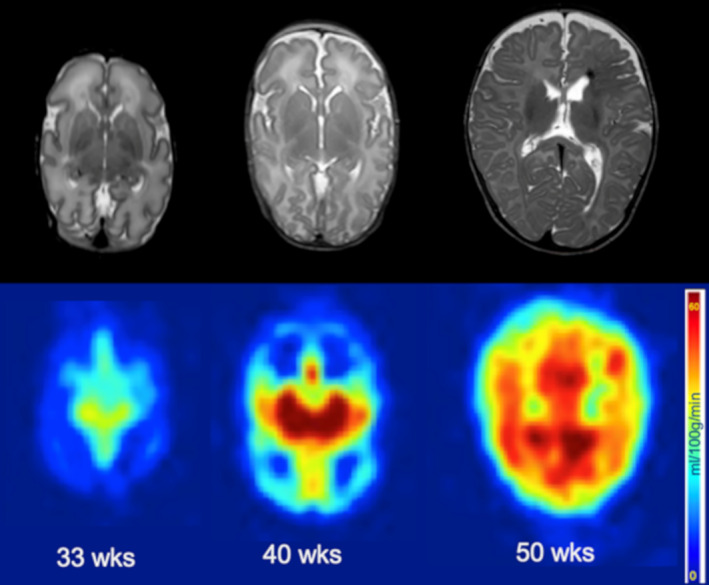
More recently observation in studies have illustrated an escalation of CBF with infants age ,that points to maturation of brain[104,105]. Maximum CBF is at the deep gray matter nuclei from the central along with occipital areas in contrast to frontal areas (figure 8),that in turn illustrated a greatest reduction in the basal ganglia in association with neuromotor results[ 80]. Lesser association with CBF values have been documented in the insula , anterior cingulate, along with auditory areas in addition to the existence of parenchymal brain damage associated with lesser global along with areas of CBF[ 107].
Provision of quantification of metabolites , is carried out by Proton Magnetic resonance spectroscopy(MRS) dependent on their variation in resonant frequency or chemical Transfer.On MRS spectra metabolites peak gets represented on a ppm scale A single voxel strategy has greater potency in contrast to, a Multi voxel strategy (like Chemical shift Imaging (CSI)with more advantageous homogeneity with regards to field, greater SNR that caused a spectral resolution, lesser Chemical shift or voxel bleeding artefacts along with lesser contamination from nonrepression of water or lipid signal. Greater strength of field aided in lesser spectral overlapping along with utilization of smaller voxel size till Bo homogeneity gets to the ideal level. At3T an adibatatic(or an event where no heat transfer takes place thoughnot mean that the temperature is constant, but rather that no heat is transferred into or out from the system),selective repeat concentration is advocated along with 1.5cm3 voxel with 128 means in neonates[108].
Alterations in the amounts of metabolites are significant, at the time of maturation of brain in particular at the time of the first postnatal year with a rapid reduction in N-acetyl Aspartate(NAA,peak at 2.02 ppm) along with creatine(Cr peak at 3.03 ppm). in addition to a rapid escalation of choline(Cho , peak at 3.2 ppm),. as well as myoinositol (mI,peak at 3.56 ppm)[109]. NAA is implicated in myelination ;its transportation out of the neurons takes place to the oligodendrocytes,where its utilization occurs for myelingeneration[ 110].Creatine is essential, for the control of energy provision in cells as well as is believed to be correlated with the neuronal cell mass.Full cholineacts as a marker for the turnover of membrane, besides myoinositol acts as a marker for glia . Greatervalues of both are illustrated in newborns.Glutamate-Glutamine (Glx, peak at 2.1-2.5 ppm) utilization takes place in the form of a marker for neuronal degradation events. At the time of generation it is implicated, in various stages involving neurogenesis as well as maturation that is inclusive of proliferation of neural progenitor cells, migration, differentiation, survival as well as synaptogenesis[ 110].Lactate(peak at 1.32 ppm)works as a marker of hypoxia or failure of cellular energy, however a minimum Lactate(peak is a normal observation in neonates[ 109].
Besides alterations in age amounts of metabolites is based on residence of brain with greater NAA as well as Cho values along with lesser Cr, mI, as well as Glx values in WM in contrast to, GM, with the maximum values of Cr, as well as Cho in cerebellum .As per the age in addition to where brain resides normative curves can get observed in the literature, however are based on the acquisition protocol [ 110].
In the neonates having a premature birth greater, Quantities of NAA, Glx as well as Cr along with mI have been illustrated in contrast to fetuses of akin age[ 111]. Preterm infants at TEA that are without any significant damages to the WM, demonstrated lesser quantities of Cr, Glx in addition to macromolecules in the WM that pointed to changes in metabolism besides protein generation[ 112], lesser Quantities of mI, that pointed to probable astrogliosis[ 113], lesser NAA/ Cho ratio in the thalamus in correlations with neuro developmental postponement, at 18mths of age[ 114], lesser NAA in addition to greater Cho quantities in cerebellum[114].The existence of cerebellar damages possessed a constant correlations with reduction in NAA, Cho along with Cr[115]. Preterm infants possessing Punctate white matter lesions further demonstrated a reduction in NAA quantities in the parietal WM, pointed to neuronal injury as well as myelin damage[ 113].
Other More Recent MRI Strategies
Various methodologies are accessible for quantitative assessment of the maturation of brain of the newborns. However, till date occasional studies have attempted contrasting the markers that are accessible as well as assessment of their supportiveness or dismissal . More recently, certain polished strategies have been pointed to give the properties of modes of maturation dependent on prior quantitative MRI techniques.Implmentation along with compartmentalization or multiparametric techniques for contrasting or incorporation of the supportive parameters.
Complicated markers of maturation
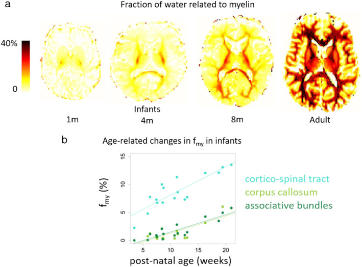
Of these recent strategies having the objective of provision of a marker of the quantity of myelin in the generating brain by the computation of the water volume fraction of water associated with myelin.This is dependent on a multcompartmental model determined dependent on the dissemination of T1 as well as T2 relaxation times in every voxel of the tissue,that needs estimation of MR signal of various acquisition setting(like variousTEs,separate TIs). Variable techniques for acquisition of data along with evaluation have been pointed for determination of parameters that attract us like ((fmy ),with varying dependence based on the model presumptions(like the chamber relaxation properties). Provision of techniques estimating dependent on the Imaging methodologies mc DESPOT(multi component driven equilibrium Single pulse observatiosa of T1 / T2 has aided in a spatio temporal pattern in agreement with earlier histological studies with regards to myelination at the time of infancy as well as toddlerhood, with the initiation in central brain areas as well as progression in a caudocranial direction[116].A data guided methodology (like independent component analysis (ICA) has aided further parcellation (like division into parcels,countable awa uncountable-equal )WMareas as per their fmy courses[117]. The courses or directions of certain areas emphasized in this manner were correlated with unique alterations in cognitive capacities.
The major limitation of this technique is the long acquisition or preprocessing time;that is an alternative technique dependent on EPI sequences was suggested,with a prior calibration step with regards to model parameters, carried out on occasional adults[118]. The full brain maps derived in infants with a 5’ protocol illustrated the propagation of myelination from central to peripheral areas (figure 9a,). In the BCP project ,a 2D MR finger printing technique have been generated for concomitant estimation of fmy along with T1 as well as T2 relaxation times[63].The initial outcomes obtained demonstrated a practically non existent fmy till 6mth age ,with smooth escalation subsequently with variations as per area (figure 9b). In contrast to T1 as well as T2 parameters apparently provision of differential however supportive sensitivityby fmy to the alterations in tissue maturation[56,116].
Other more recent combined techniques pointed to determine the the - g-ratio (like the ratio of the axons diameter with the outer fibers diameter inclusive of the myelin sheath ) dependent on diffusion along with myelin associated estimates(with utilization of,like NODDI indices,MTR or fmy estimates)This marker is anticipated to result in reduction in the WM with the myelination event in view of it being found in preterm infants that have been going via Imaging at 30 as well as 40 w PMA [119], as well as In young children [120]. Provision of appropriate knowledge via this index with regards to effectiveness, of shift of Neural information in addition to the conduction velocity of WM pathways. Nevertheless, the harmonious nature of these variation of markers in association with maturation modes , has requirements of evaluation of variable generational durations.
Occasional studies have initated the evaluation of this problem. Contrasting of T1,MTR along with DTI parameters In infants having undergone scanning at the time of the preterm duration(28-32w PMA) as well asTEA illustrated that these maps possess unique contrasts with variation amongst, brain areas along with over ages [51]. In contrast to other parameters the pattern of MTR of variable alterations in areas amongst, the preterm age along with TEA.Voxel dependent evaluation along with contrasting of these parameters emphasized further the pattern of lamination in the wall of the cerebrum along with pointed to variable modes of maturation in the brain chambers(like subplate, intermediate zone)[67]. In case of preterm infants amongst 27 as well as 58w PMA NODDI, T2, as well as characteristics of fmy were further contrasted i in variable brain areas [121].As per this study it was demonstrated that in the thalamus diffusion age associated alteration are not present only secondary to myelination ,whereas diffusion along with T2 alteration were correlated with posterior WM (possibly secondary to axonal along with glial proliferation) however are not dependent on myelin water quantity.
Multi parameteric strategies
Other strategies have been suggested more recently for provision of the incorporation of the harmonious knowledge by MRI parameters (T1 as well as T2 relaxation times, anisotropy, along with diffusivities of DTI) in infants amongst 1 as well as 5mths of postnatal age with the objective of taking into account alterations associated with different maturational modes together(like alterations in cell along with membranedensity in water along with iron quantity in association with the generation of dendritic arborisation, synaptogenesis, myelination of fibers,etc). Initially a strategy was taking into account for grouping brain voxels dependent on their akin characteristics ,with the application of clustering algorithm(alias a process or set of rules to be followed in calculations) to combining these indices [68,9].This aided in the classification of cortical areas as per their maturation, without any posit a priori(alias proceeds from theoretical deduction rather than from observation or experience) or their anatomical placement[55]. The resultant maps illustrated variable maturation pattern of cortical areas at the unique level in addition to propagation across the infants group as per age .This corroborated the early maturation of primary sensorimotor areas with subsequently neighbouring unimodal associative areas, along with finally higher order associative areas (figure10a). T1 as well as T2 axial diffusivity(λ// )then was apparently the maximum dependent parameter for evaluation of the properties of GM across this generating duration .Akin strategy was applicable to the WM voxels emphasized the way progression of maturationoccurred from the centre of the brain to the periphery[54].
Additionally, a maturation spacing (dependent on the Malanobis distance )got determined in a set of bundles of WM,by contrasting T1, T2, as well as DTI diffusivities in infants from that of group of adults[53].This illustrated greater association amongst, bundles in contrast to univariate strategies , along with aided in quantification of their apparent postponement, of maturation .The outcomes corroborated the potent alterations in the postnatal yr along with the asynchrony with regards to maturation over the bundles, Noticeably with early maturation of the corticospinal tract ,fornix, in addition to spinothalamictract as well as optic radiations along with postponement, of maturation of associative bundles (like arcuate, as well as superior longitudinal fasiculus)(figure10b).
In toto with these studies provision of gadgets along with microstructural markers which might cause more advantageous reflection of the complicated along with overlapping patterns of maturation modes in the WM as well as GM tissues right via generation . Nevertheless, greater works are required for ascertaining the translation of these techniques into clinical scenario.
Functional MRI
At the time of early infancy structural maturational modes as determined with the MRI methodologies detailed earlier are correlated with the functional generation of the brain aiding infants for acquisition of sensorimotor along with cognitive capacity of their perception of microenvironment, experiences as well as learning.
Actually, despite immature at birth the brain demonstrated an early architecture[17]. However significant plasticity events might possess a role along with cause a modification of the early generated architecture specifically under the restraint of variation of disruption(like perinatal damage).
Generational specificity along with Technological hurdles of MRI
Mapping of the functional networks utilization represents the commonest technique of fMRI. However,this is tough in the case of infants [122].Akin to the prior MRI methodologies, short protocols might possess preference in the contex alm . Actually,head movements constitute significant problems as well in view of variable etiologes of this behaviour, apart from variations amongst, group of infants(classical or at risk for generation of non classical generation that result in artifactual confounders in fMRI studies[123]. More recently a custom fashioned MR coil has been pointed to result in optimization of the temporal SNR as per the size of the head of newborn[124] along with in the dHCP Project utilization of a custom fashioned 32 channel neonatal particular head coil[28]. Maximization of the sensitivity for the BOLD(blood oxygen level dependent responses )in brain activity with utilization of ideal TE in association with T2* properties of the generating brain (greater T2* in newborn along with infants in contrast to adults in view of greater water quantities along with lesser lipid quantities).For3T fMRI of full term newborns,a TE of >50msec has been advocated for determination of significant stimulation associated BOLD alterations[125].
With regards to post processing of fMRI outcomes, variable steps need to be taken into account for maximization of the dependability along with accuracy of functional activation maps .The dHCP resting state fMRI(rs- fMRI) re processing pipeline has been expanded for or the assessment of stimulation- response fMRI images[126].Implementation in addition to deformation along with rectification,ICA dependent denoising as well as haemodynamic modeling resulted in a considerable escalation of sensitivity as well as specificity of functional maps[126]. Actually, the BOLD response properties(like time duration,amplitude ) evolution at the time of generation. Determination of a precise model of the haemodynamic response function(HRF)in the population of patients targeted is needed .Like with a stimulation prototype along with a process associated fashion,the properties of HRF waveform has been studied in the preterm newborns at 32-39wPMA, at TEA(38-44wPMA)along with in adults in addition to systematic maturational tendencies were seen ;the amplitude of the major positive peak escalated with age whereas a reduction of time was seen[127].
In contrast to adults, BOLD responses with positive /negative peaks have been documented in neonates along with infants[128],that has evoked a debate on the generational alterations in CBF along with oxygen consumed subsequent to neural activity. Actually, the neurovascular coupling(mode associating the temporary neural activity to the following alterations in blood flow ) in addition to automatic controlling system are apparently different in the generating brain in contrast to adult[129].The properties of generational evolution in cortical blood flow was assessed in a systematic study in neonatal rats[130].P12 rats(equal to the human newborns) demonstrated an inverted haemodynamic response(negative BOLD)with the consumption of early oxygen along with postponement, of constriction of pial arteries.These responses differed with the stimulus stimulation of systemic blood pressure(BP),that resulted in cortical hyperaemia.In case of rats that were older maturation of haemodynamic response took place with the generation of a starting phase of hyperaemia(-positive BOLD)which in the end had a masking action on oxygen consumed, along with balance of vasoconstriction towards adult stage[130].These kinds of dynamic alterations might reason out the fluctuations in BOLD responses detailed in prior fMRI studies on newborns as well as infants.
More recently fMRI at the time of stimulation archetypes have been utilized for assessment of the generating brain events sensorimotor along with cognitive knowledge (sensorimotor visual, along with language stimuli)
i)Sensorimotor systems-Somatosensory Insight along with motor activities are the initial ones that the fetuses, experience with in the womb The evaluation of sensorimotor networks are dependent on the passivemotor along with tactile stimulation archetypes figure 11a). Brain responses subsequent to induced wrist movements have been isolated in the opposite primary Somatosensory along with motor cortices in case of pre term newborn that are as small as 30wPMA[131]. fMRI further demonstrated that a rough somatotopic organization, of the primary cortices is in place from 32wPMA,with brain areas whose activation occurs by stimulation of the wrists,ankles as well as mouth[132]. With regards to evaluation of the generating brain events affective touch, another group determined brain responses to gentle skin stroking in full term newborns that were under 1mth age as well as illustrated activations in 2 areas that were implicated in the mature brain(post central gyrus along with posterior insular cortices) [133].These studies pointed to an early functional organization, of the sensorimotor networks from the preterm duration.


ii)Sense along with smell systems ; smell is the2nd sense which generates in fetuses, in utero . Subsequent to birth ,it has been anticipated to possess a key part in behavioural adaptations crosstalk along with bonding events which occur amongst the newborn along with mother. More recently a fMRI evaluated in newborns illustrated adult like cortical areas(inclusive ofpiriform,orbitofrontal along with insula) get activated by the discernment of olfactory as well as trigeminal to new odorants [172](figure11b)., pointed to an early specialization of the smell brain networks .
iii)Visual systems vision is amongst, the first sensory functions that generates in humans. Visual stimulations ‘’in utero ‘,are pretty restricted however spontaneous retinal activity aids the visual pathway to initate function, as well as specialize in the third trimesters of GA.A hurricane of visual stimuli get triggered by birth which would result in induction of escalation of cerebal activity in addition to a sequelae of modes of maturation .Their is difficulty in implementation of fMRI visual protocols in infants,thus the initial studies were performed in sedated patients along with illustrated negative followed by positive BOLD responses in newborn along with infants (figure11c)[128]. Nevertheless, recently 2 groups attained success in evaluation of awake infants. dependent on flow vs random motion stimuli,the main cortical areas attributable to visual motion insight were demonstrated to be active in 2mth old infants , in addition to unique visual inputs to primary (VI) as well as temporo occipital(VS/MT+)areas appear to be existent early[135]. Inspite of variation in the profiles to responses along with activity paradigms,the extrastriate visual was demonstrated to display an early adult like specialization along withspatial organization, for visualcategories(like faces,scenes )in 4-6 mth old infants[136].As per these studies,a few mths following birth the generating brain possesses the capacity of high level visual processing despite the refinement of underlying networks.
iv)Auditory / language systems; The maturation of auditory systems spans over longer duration in contrast to visual systems from pregnancy to childhood .The brain networks committed to auditory along with language systems processing have been assessed by various groups with fMRI . In 3mths old infants speech stimuli possessed capacity of stimulation of activity in left lateralized brain areas (inclusive of superior temporal, as well as angular gyri) [137] (figure11d).Like in adults ,perisylvian areas demonstrated variable speeds of activation (with maximum rapid response intricate to Heschl’s gyrus ) along with variation in sensitivities to repetition of sentences(with escalation of activity in inferior frontal Broca’s areas pointed to early implication in verbal memory)[138]. In contrast to biological non speech sounds,the temporal, area becomes activated amongst, 1 as well as 4 mths of age[139]. Conversely in full term newborns, listening to music resulted in stimulation of right hemispheric activation of the primary along with higher order auditory cortical areas[140]. In contrast to full term newborn, preterm infants at TEA(38-41wPMA) illustrated lesser posterior thalamic activation to linguistic stimuli however akin bilateral activation in superior temporal, supramarginal, inferior frontal gyr[i141]. At the time of early age(29-34wPMA) superior temporal as well as supramarginal activation have been further seen with left predominance,whereas at a later stage(44-45wPMA) there is refinement of paradigm of activity of bilateral superior temporal as well as left lateralized supramarginal activation.From TEA these brain responses appeared partially associated to the Neuropsychological results in preterm infants[141]. In total these studies pointed to an early lateralized specialization for speech as well as music processing which would get manipulated by the experienced milieu at the time of preterm duration ,
With the advancements in these task dependent fMRI studies in newborns are basal. However their implementation is tough In particular with the situations of installation in the MRI scanner the noise with regards to acquisition along with sensitivity to motion cause reduction of the probability that the baby is awake as well as calm at the time of the protocol.In clinical scenario thus the probability of these studies is reduced without any major advancements in these variable domains.
Mapping Resting state networks
The other methodology that is attractive for the evaluation of the generating brain in newborns is rs- fMRI .It aids in mapping areas possessing ‘’functional connection’’,like areas that illustrated spontaneous as well as logical BOLD signal variations across time .Generally variations at low frequencies (like 0.01-0.08Hz that were complementary to time durations of 12.5-100sec)get targeted. Nevertheless, evaluation offunctional connection’’ in high frequency bands have been suggested as well along with that of dynamic (rather than at stand still) ’functional connection’’[144].The major benefit of rs- fMRI in newborns is the acquisition of results without task. However its Impications are variable technological hurdles hurdles as per prior publications[144].As per the assessment of results,a first technique is ROI- dependent, besides provision of voxelwise association evaluation of the full brain.The assessment of outcomes of these is easy, however they are remarkably based on the anatomical definitions as per the ROI. Identification of the unique areas dependent on the progression, of an atlas where one can subdivide data into parcels. However need for an age matched atlas is feasible in addition to correct registration amongst, the group[144]. One more strategy is data based ICA(figure12), that aids ruling out noise constitutents, in future evaluation.
More recently studies have illustrated that the newborn illustrates a complicated resting state functional architecture with generational alterations in interhemispheric connectivity in the preterm duration[81]. Assessment of connectome gradients in full term newborns has illustrated variation amongst, unimodal as well as trans modal areas that pointed to adult like organization when under generation[143], Networks existent in visual, sensorimotor along with auditory areas are existent already subsequent to birth[144], however their volume besides power of activation escalation, at the time of 2yrs just of age[145]. Amongst 4 as well as 9mths of age an akin network localization is present, however reduction inthe connectivity power in local networks in addition to escalation in more spaced networks[146]. With regards to, higher order cognitive ,ie an immature default mode network(proto DMN)( dependent on ventomedial prefrontal, posterior cingulate,medial temporal, areas,pre cuneus in addition to angular gyrus)appear at the age of 1 yr[147]. Central executive network(depending on dorsolateral prefrontal, posterior parietal areas )is further seen in full term newborns[144],despite escalation of connectivity takes place at the time of generation. In the context of salience network (depending on anterior insula as well as dorsal anterior cingulate area) local specialization as well as connectivity escalation occurs at an astonishing pace at the time of initial 1st 2yrs of age[148].
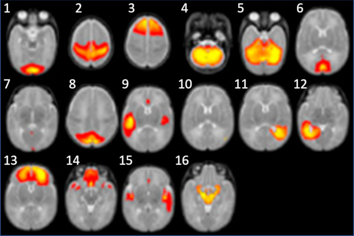
Certain studies in newborns have started contrasting the generating structural along with functional connectivity in paradigm whose provision is made by rs- fMRI along with diffusion MRI.The modes that associate these determinants is not clear till date in adults;the power of structural connections apparently result in anticipation of the power of functional contacts,while powerful functional contacts are existent amongst, areas not processing robust functional contacts amongst areas with no direct functional contacts[149]. Certain studies have further demonstrated that structural akinness as well as non akin amongst, the structure as well as functional connectivity paradigms dependent on the system with more overlap in sensory areas along with pathway in contrast to higher order association types[75,81].This might be secondary to variations in maturation, amongst systems that result in biases with regards to methodologies (like the more dependable tractography reconstructions of mature structural communications connections (like sensorimotor in contrast to \to associative ones[81,143]. Conversely, associative areas might have.demonstrated functional incorporation inspite of a prior illustrated structural connections . Prematurity might influence the functional connectivity amongst the resting-state networks. In contrast to full-term newborns, preterm infants at TEA demonstrated lesser complicated intrinsic activity, that was lesser severe interhemispheric and thalamocortical connectivity, in addition to lesser connectivity amongst thalamus as well as and the salience network.[150]. Furthermore, they illustrated that coupling of a circuit of three network modules They demonstrated reduction in crossconnection by the salience network.[151] . These modules comprised of 1) medial superior frontal, auditory, along with a sensorimotor networks; 2) orbitofrontal, posterior cingulate areas , precuneus, visual along with left posterior temporal networks; along with 3) thalamus precuneus, as well as right posterior temporal networks. Environmental enrichment via music exposition resulted in escalation of the functional [connectivity in this particular circuit[151].
This study further pointed to an original strategy for assessment of functional connectivity on nonparametric dependent onevaluation determinators of accordance in addition discordancefor escalation of the sensitivity and severity of detection of group actions ,It presumed that extreme processes of the BOLD fluctuations pointed to significant activations or deactivations of the ROI, whereas spurious fluctuations are thought of noise. In all, the rs-fMRI strategy is attractive for acquiring insight in the context of the generation of functional networks in newborns,in particular when the results can be contrasted with the ones whose provision was by complementary MRI Methodologies for assessment of structural maturation..
Conclusionsandfutureperspectives:
More recently wide types of innovative techniques have been pointed to along with implemention in aiding the MRI study of the generating brain in vivo in newborns in addition to infants. These variable strategies aid in the escalation of the appropriate assessment of multiple neurodevelopmental modes, from morphological to microstructural alterations, visualized in both GM as well as WM, along with metabolic as well as functional alterations. Nevertheless studies of the generating brain remain restricted in various aspects,with the knowledge given that intrinsic technological in addition to experimental challenges,.It is tough for advocating in general
without the information with regards to query being posed, the clinical or research context, along with and the accessible acquisition time in the context protocol in newborns. Quality control procedures are necessary, to ascertain the biological veracity of MRI findings observations. Hence, numerous hurdles continue to be tackled for assessment, in the most automatic in addition to dependable method feasible cohorts with a large number of subjects of variable ages, or groups of newborns with variable early pathologies. High-field MRI (ie, 7TMRI) would also be an exciting as well as attractive perspective for mapping, with high spatial resolution, fine structures information, or microlesions not seen with 3T MRI. This is a topic that has become debatable with certain researchers and clinical groups have started addressing, however which result in stimulation of additional restraints along with hurdles. Knowledge.
Another necessary perspective at present bothers multimodal strategies with the objective of
combination of brain MRI determinants with clinical in addition to behavioral markers, or electrophysiological indices like those whose provision is by electroencephalography(EEG)[75]. For example, an escalation of, activity in the first postnatal days in preterm newborns (as determined with EEG signalpeak-to-peak amplitude and “spontaneous activity transients”) has been related to a faster growth of the brain along with subcortical GM at the time of following weeks until TEA[152].In classical infants, research studies have further pointed to important association amongst with MRI microstructural indices of WM maturation as well as EEG functional measures (ie, response latency and speed) for the visual system[153 ].But such associations apparently possess
lesser clarity in the context the auditory system,[71] pointing to a greater complicated crosstalk, modes , in which the environment, , might play a greater significant part.Acquiring insight in the context of generational asynchronies withregard to the early sensory in addition to incorporation capacities of infants continues to be an exciting question . MRI possesses a critical part play here, along with to greater advantage with regard to properties in addition to diagnosing neurodevelopmental pathologies from pre- or perinatal origins. Since it can be attained well before possessing r knowledge in the context of child’s behavioral along with clinical results MRI becomes an unavoidable assessment technique for evaluation of the effectiveness of early neuroprotective or neuroregenerative therapeutic treatments or strategies for correction with the objective of avoidance of long-term disabilities of children.
Dear Editorial Team, Clinical Medical Reviews and Reports. My experience with the journal was highly positive. The peer-review process was rigorous, constructive, and completed in a timely manner. The reviewers provided valuable comments that helped improve the quality and clarity of our manuscript. The editorial office was professional, responsive, and supportive throughout all stages of the publication process. Communication was clear and efficient, and any questions were addressed promptly. Overall, I found the journal to maintain high scientific standards and an excellent publication workflow. I would be pleased to consider submitting future work to this journal. Best wishes from, Elena Popa.

It was my pleasure to submit my testimonial concerning the Reviewer Board of our Scientific Journal “Brain and Neurological Disorders”. The Reviewers focused on some modifications and their contribution was helpful. The ladies of our Editorial Office were also supported my efforts. It was my honor to have such a co-operation and I am looking forward for more collaboration.

Dear Grace Pierce, Editorial Coordinator of Journal of Clinical Research and Reports, Thank you for the speedy and efficient peer review process. I appreciate the fact that your peer reviewers do not take months to respond like with some other journals. I would also like to thank the editorial office for responding quickly to my questions. It is an excellent journal. I plan to submit more manuscripts in the future. Best wishes from, Robert W. McGee

Dear Grace Pierce, Editorial Coordinator of Journal of Clinical Research and Reports, Working with you and your team on our recent publication in JCRR has been a truly wonderful and enjoyable experience. The responses were prompt, and the reviewers were patient, constructive, and highly professional. One reviewer in particular gave me the feeling that a professor was carefully reading and commenting on my coursework, which was deeply touching. The entire process was straightforward and hassle‑free, with no tedious online forms to complete. I highly recommend this journal. Best wishes from, DR Aibing Rao, Head of R&D

I Appreciate the Opportunity to Share my Experience with the Journal of Clinical Research and Reports. The peer review process was timely and constructive, and the feedback provided helped improve the quality of our manuscript. The editorial office was professional, responsive, and supportive throughout the process, ensuring smooth communication and efficient handling of the submission. Overall, it was a positive experience collaborating with your team.

Dear Mercy Grace, Editorial Coordinator of Obstetrics Gynecology and Reproductive Sciences, We would like to express our gratitude for your help at all stages of publishing and editing the article. The editors of the magazine answer all the necessary questions and help at every stage. We will definitely continue to cooperate and publish other works in the Obstetrics Gynecology and Reproductive Sciences! Best wishes from, Alla Konstantinovna Politova,
