
Case Report | DOI: https://doi.org/10.31579/2690-4861/187
Critical Care Unit, Kafr El-Bateekh Central Hospital, Damietta Health Affairs, Egyptian Ministry of Health (MOH), Damietta, Egypt.
*Corresponding Author: Yasser Mohammed Hassanain Elsayed, Critical Care Unit, Kafr El-Bateekh Central Hospital, Damietta Health Affairs, Egyptian Ministry of Health (MOH), Damietta, Egypt.
Citation: Y M H Elsayed. (2022). Anaesthetic and perioperative management of elderly female with post covid ARDS with comorbities undergoing emergency Laparotomy for obstructed umbilical hernia. International Journal of Clinical Case Reports and Reviews. 10(3); DOI: 10.31579/2690-4861/187
Copyright: © 2022 Yasser Mohammed Hassanain Elsayed, This is an open access article distributed under the Creative Commons Attributiosn License, which permits unrestricted use, distribution, and reproduction in any medium, provided the original work is properly cited.
Received: 22 October 2021 | Accepted: 11 January 2022 | Published: 19 January 2022
Keywords: Covid-19; pneumonia; stroke; qtc-interval prolongation; fragmentation of the qrs-complex; connected aircraft squadron electrocardiographic sign (yasser’s sign)
Rationale: COVID-19 infection is a wide-world pandemic serious highly infectious multisystem disease. Neurological, cardiovascular, hepatic, and renal systems are commonly involved. Connected aircraft squadron electrocardiographic sign (Yasser’s sign) is a novel electrocardiographic sign which a relevant to T-wave changes and respiratory rate. This sign is a strong guide for tachypnea. QTc-interval prolongation is a serious signal for torsades de pointes, fatal ventricular tachyarrhythmias, and sudden death. Previously, fragmentation of the QRS-complex had already been considered a marker for structural heart diseases (SHD) triggering cardiac hypertrophy.
Patient concerns: An 88-year-old, carpenter, smoker, married Egyptian male patient was admitted to the critical care unit with cerebrovascular stroke, cardiac involvement, and COVID-19 pneumonia.
Diagnosis: COVID-19 pneumonia with acute neurological events and cardiovascular involvement.
Interventions: Electrocardiography, oxygenation, non-contrasted chest CT, and brain CT.
Outcomes: Good response and better outcomes despite the presence of several remarkable risk factors were the results.
Lessons: The clinical and electrocardiographic response after using anti-COVID19 measures the signifying its role and suggest the diagnosis of COVID19 infection. The presence of COVID-19 pneumonia, stroke, QTc-interval prolongation, fragmentation of the QRS-complex, hypocalcemia, Connected aircraft squadron electrocardiographic sign (Yasser’s sign) with severe fatigue, elderly, and cigarette smoking are prognostic factors for the severity of the disease.
COVID-19: Coronavirus disease 2019
ECG: Electrocardiogram
ICU: Intensive care unit
O2: Oxygen
QTC: corrected QT interval
SGOT: Serum glutamic-oxaloacetic transaminase
SGPT: Serum glutamic-pyruvic transaminase
TdP; torsades de pointes
VR: Ventricular rate
Acute ischemic cerebrovascular accident (CVA) may happen in cases of coronavirus disease 2019 (COVID-19). The risk factors involving both in-hospital events and outcomes are still not well-been studied in large cohorts [1]. Although there is a relationship between QT-interval duration and the risk of torsades de pointes (TdP) is still not fully understood, a corrected QT interval (QTC) of >500ms [2] or an increase in the QTC of >60ms [3] is mostly index for a high risk of TdP. However, QT prolongation is an inducer marker for the developing TdP. Screen for other risk factors for QT prolongation, including drug interactions, electrolyte disorders such as hypokalaemia, and renal dysfunction are is pivotal in management. ECGs should be performed in all patients with symptoms of arrhythmia and periodically in patients at high risk of QT prolongation/TdP4. Correct modifiable risk factors areessential [4]. The term ‘fragmentation of the QRS-complex” or “fragmented QRS” (fQRS) point to the existence of high-frequency potentials (spikes) in the QRS-complex [5]. The expression was first described in 1973 in reporting of an experimental study on canine hearts where coronary artery obstruction motivated the occurrence of fragmented electrograms as a source of reentrant activity [6]. Formerly, fQRS had already been considered a marker for structural heart diseases (SHD) triggering biventricular hypertrophy [7]. The presence of fQRS can be induced by any condition interrupting with the normally homogeneous depolarisation status in the myocardium and yielding regional conduction decelerating such as ischemia, scar, fibrosis, myofiber disarray, inflammation, and microvascular abnormality [5]. Das et al. revealed that there was a good correlation between fQRS and the presence of myocardial scar in patients with ischemic heart disease (IHD) that appeared by single-photon emission tomography (SPET) [8]. Connected aircraft squadron electrocardiographic sign is a new strong index for monitoring and follows up the tachypneic patients with specific T- waves changes in special leads in several cardiorespiratory patients [9]. Three types were described [1]. Type I: comprises only eighteen patients of investigated tachypnea with T-wave changes among both I and II leads, and with aVL and aVF leads [2]. Type II: comprises four patients of tachypnea with T-wave changes only between aVL and aVF leads [3]. Type III: comprises two patients of tachypnea with T-wave changes only between V1 and V2 leads [9]. There are four reported morphological degrees for the sign: 1. first degree: the presence of sign configuration (shared) [2]. Second degree: the presence of approximation between the target inverted and an upright T-wave [3]. Third degree: the presence of connecting the target inverted and an upright T-wave. 4. Fourth degree: the presence of gap interlacing both the target inverted and an upright T-wave [9] .
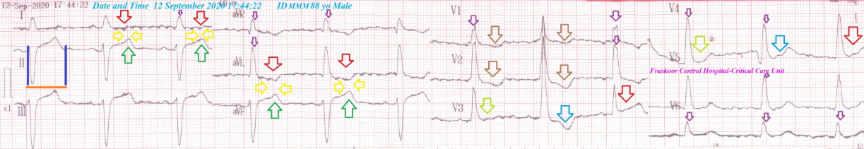
An 88-year-old married, Carpenter, smoker, Egyptian, male patient was admitted to the intensive care unit (ICU) with left side weakness and dizziness. Generalized body aches and fatigue were the associated symptoms. He gave a history of fever, cough, and generalized body aches 4 days ago. The patient is a currently heavy smoker (at least 20 cigarettes for about 40 years). He denied a history of cardiovascular diseases, the same attack, drugs, or any other special habits. There was a recent positive history for contact with a COVID-19 confirmed patient. Informed consent was taken. Upon general physical examination; generally, the patient was in awareness, alert, and wakeful, with GCS; 15 regular heart rate of 60 bpm, blood pressure of 110/70 mmHg, respiratory rate of 10 bpm, the temperature of 36 °C, and pulse oximeter of O2 saturation of 91%. Deep reflexes, sensation, and gait were normal. No more relevant clinical data were noted during the clinical examination. Urgent initial ECG tracing was done on the presentation in the ICU showing type II, grade I Connected aircraft squadron electrocardiographic sign (Yasser’s sign), dominant R-waves in V1 and V2 with T-wave inversions, QTc-interval prolongation, a Wavy triple sign appears in V3 and V4 leads, and QRS-fragmentations (Figure 1). O2 inhalation (100%, by nasal cannula, 5L/min) was given. The patient was maintain treated with cefotaxime; (1000 mg IV every 8hours), azithromycin (500 mg PO single daily dose), oseltamivir (75 mg PO twice daily only for 5 days), and paracetamol (500 mg IV every 8 hours as needed). SC enoxaparin 40 mg twice daily), aspirin tablet (75 mg, once daily), clopidogrel tablet (75 mg, once daily), and hydrocortisone sodium succinate (100 mg IV every 12 hours) were added. The patient was daily monitored for temperature, pulse, blood pressure, and O2 saturation. The initial complete blood count (CBC); Hb was 10.7 g/dl, RBCs; 3.95*103/mm3, WBCs; 11.2*103/mm3 (Neutrophils; 84.6 %, Lymphocytes: 12.4%, Monocytes; 2%, Eosinophils; 1% and Basophils 0%), Platelets; 158*103/mm3. Serum creatinine was normal (0.9 mg/dl) and blood urea was normal (29 mg/dl). RBS was normal (179 mg/dl). Ionized calcium was slightly low (0.78 mmol/L), plasma sodium was normal (141 mmol/L), and serum potassium was normal (4.6 mmol/L). SGPT was normal (18 U/L), SGOT was normal (24 U/L). Total bilirubin was normal (0.7 mg/dl). D-dimer was normal (302 ng/ml). CRP was high (30.4 g/dl). Ferritin was high (387 ng/ml). LDH was high (275 U/L). The troponin test was negative (0.1 ng/L). Chest CT without contrast was done within 5 days of the presentation showing bilateral patchy ground-glass pulmonary consolidation in the peripheral, basal, and posterior segments (Figure 2A and 2B). A brain CT scan was done within 4 days of the presentation showing evidence of matched brain atrophic senile changes with chronic ischemic white matter changes. (Figure 2C). COVID-19 pneumonia with neurological and cardiovascular involvement was the most probable diagnosis. The patient was discharged on the fifth day after clinical and electrocardiographic improvement. The patient was continued: aspirin tablet (75 mg, once daily) and clopidogrel tablet (75 mg, once daily). The patient was advised for cardiovascular, pulmonary, infectious, and neurological diseases follow-up.

Overview
A 88-year-old married, Carpenter, smoker, Egyptian male patient was admitted to the ICU with cerebrovascular stroke, cardiac involvement, and COVID-19 pneumonia.
The primary objective for my case study was the presence of a patient who presented with cerebrovascular stroke, cardiac involvement, and COVID-19 pneumonia in the ICU.
The secondary objective for my case study was the question of; How did you manage the case at home?
The presence of direct contact to confirmed the COVID-19 case, and bilateral ground-glass consolidation, lymphopenia, neutrophlilia with higher activity indices such as CRP, s-ferritin, and LDH will strengthen the COVID-19 diagnosis.
The presence of elderly, pneumonia, stroke, heavy cigarette smoking, QTc-interval prolongation, and QRS-fragmentation are aconstellation of serious risk factors.
The presence of fragmentation of the QRS-complex is considered another risk.
There is a type I, grade I Connected aircraft squadron electrocardiographic sign (Yasser’s sign) that may be an indicator for severe fatigue [9].
The existence of stroke with brain CT scan evidence of chronic ischemic white matter changes is mostly aggravated by COVID-19 infection.
I can’t compare the current case with similar conditions. There are no similar or known cases with the same management for near comparison.
The only limitations of the current study are the absence of serial ECG tracings at the time of hospitalization.
The clinical and electrocardiographic response after using anti-COVID19 measures the signifying its role and suggest the diagnosis of COVID19 infection.
The presence of COVID-19 pneumonia, stroke, QTc-interval prolongation, hypocalcemia, connected aircraft squadron electrocardiographic sign (Yasser’s sign) with severe fatigue, elderly, and cigarette smoking are prognostic factors for the severity of the disease.
I wish to thank the team of nurses in the critical care unit in Faraskour Central Hospital who make extra-ECG copies for helping me. Also, I want to thanks my wife to save time and improving the conditions for supporting me.
Conflicts of interest
There are no conflicts of interest.
Dear Editorial Team, Clinical Medical Reviews and Reports. My experience with the journal was highly positive. The peer-review process was rigorous, constructive, and completed in a timely manner. The reviewers provided valuable comments that helped improve the quality and clarity of our manuscript. The editorial office was professional, responsive, and supportive throughout all stages of the publication process. Communication was clear and efficient, and any questions were addressed promptly. Overall, I found the journal to maintain high scientific standards and an excellent publication workflow. I would be pleased to consider submitting future work to this journal. Best wishes from, Elena Popa.

It was my pleasure to submit my testimonial concerning the Reviewer Board of our Scientific Journal “Brain and Neurological Disorders”. The Reviewers focused on some modifications and their contribution was helpful. The ladies of our Editorial Office were also supported my efforts. It was my honor to have such a co-operation and I am looking forward for more collaboration.

Dear Grace Pierce, Editorial Coordinator of Journal of Clinical Research and Reports, Thank you for the speedy and efficient peer review process. I appreciate the fact that your peer reviewers do not take months to respond like with some other journals. I would also like to thank the editorial office for responding quickly to my questions. It is an excellent journal. I plan to submit more manuscripts in the future. Best wishes from, Robert W. McGee

Dear Grace Pierce, Editorial Coordinator of Journal of Clinical Research and Reports, Working with you and your team on our recent publication in JCRR has been a truly wonderful and enjoyable experience. The responses were prompt, and the reviewers were patient, constructive, and highly professional. One reviewer in particular gave me the feeling that a professor was carefully reading and commenting on my coursework, which was deeply touching. The entire process was straightforward and hassle‑free, with no tedious online forms to complete. I highly recommend this journal. Best wishes from, DR Aibing Rao, Head of R&D

I Appreciate the Opportunity to Share my Experience with the Journal of Clinical Research and Reports. The peer review process was timely and constructive, and the feedback provided helped improve the quality of our manuscript. The editorial office was professional, responsive, and supportive throughout the process, ensuring smooth communication and efficient handling of the submission. Overall, it was a positive experience collaborating with your team.

Dear Mercy Grace, Editorial Coordinator of Obstetrics Gynecology and Reproductive Sciences, We would like to express our gratitude for your help at all stages of publishing and editing the article. The editors of the magazine answer all the necessary questions and help at every stage. We will definitely continue to cooperate and publish other works in the Obstetrics Gynecology and Reproductive Sciences! Best wishes from, Alla Konstantinovna Politova,
