
Research Article | DOI: https://doi.org/10.31579/2578-8868/059
*Corresponding Author: Thiago Bassi, Research and Development at Lungpacer Medical Inc. Vancouver, Canada.
Citation: Bassi T, Rohrs E, Fernandez K, Ornowska R, Reynolds S, An Easier Method to Analyze Stereologically the Pig’s Hippocampus. J Neuroscience and Neurological Surgery. 4(3): Doi:10.31579/2578-8868/059
Copyright: ©2019. Thiago Bassi . This is an open-access article distributed under the terms of the Creative Commons Attribution License, which permits unrestricted use, distribution, and reproduction in any medium, provided the original author and source are credited.
Received: 17 July 2019 | Accepted: 23 July 2019 | Published: 25 July 2019
Keywords: neuroscience; stereological method; hippocampus; pigs
Most neuro-pathophysiological research involving the hippocampus uses rodents as a nonhuman model, due to the extensive experimental descriptive literature and favorable cost. The findings from the rodent hippocampus may not be generalizable to humans, though, as ontogenetically they have different hippocampal components. While the rodent model has these limitations, the use of nonhuman primates is often not feasible due to economic and ethical considerations. Pigs are a translational alternative due to the anatomic similarity of the hippocampus in humans and pigs. Materials and Methods: Eight pigs’ brains were harvested and then analyzed according to our previously established technique. Five brains were frozen and three were stored in formalin. All eight brains were then sent to an independent histology service, where they were sectioned according to the methodology established by Holm 1994. The slabs were 10 µm with 2.5 cm2 of hippocampus cross-sectional area. Results: The mean total hippocampus volume was 892.84 mm3 ± 198.91 mm3 using Holm’s methodology. The mean number of cells per sample (20X magnification settings) was 9996.75, using automated ImageJ cell counting. Discussion: In this study, the counts of hippocampus cells were divided into two regions of interest: CA1 and CA3. Our results show that the mean number of hippocampus cells observed was 5.75 million and 2.25 million, in the CA1 and CA3 regions respectively. Holm reported 4.12 million cells in the CA1 region and 1.51 million cells in the CA3 region. The results presented here indicate the CA1 and CA3 cell percentages being 23% and 9% respectively, which are similar to the percentages reported by Holm (21% and 12%). Conclusion: These results corroborate previous findings and demonstrate a novel and cost-effective way to study the hippocampus of pigs in translational neurological research.
Most neuro-pathophysiological research involving the hippocampus uses rodents as a nonhuman model, due to the extensive experimental descriptive literature and favorable cost. The findings from the rodent hippocampus may not be generalizable to humans, though, as ontogenetically they have different hippocampal components. Moreover, the hippocampus proportions and architectonics are significantly different between rodents and humans. While the rodent model has these limitations, the use of nonhuman primates is often not feasible due to economic and ethical considerations. Pigs are a translational alternative due to the anatomic similarity of the hippocampus in humans and pigs. In addition, pigs are a well-accepted animal research model and have lower cost when compared to nonhuman primates.
Human-size pigs have been used in research due to their anatomical and physiological similarity to humans. [1,2] The pig’s brain has a gyrencephalus similar to humans, which facilitates surgical procedures and interventions. Pigs are genetically relatively homogeneous, similar to inbred laboratory rats, leading to a minimal interindividual variation.
A publication by Holm in 1994 provided a quantitative description of fundamental structural parameters, regional volumes and neuron numbers in the hippocampus of the domestic pig [3]. The volumetric and numerical data presented by Holm provided a unique opportunity to evaluate structural differences in homologous hippocampus areas between pigs and humans, and obtain a better understanding of the functional consequences of the differences in size. Despite this strong foundation, the pig hippocampus has not been clearly established as the translational model of choice for neurological studies. In 2016, van Dijk published a systematic review comparing the hippocampus in 18 different species, where one pig study was reported [4].
The hippocampus is well recognized as the hub of neuroplasticity and neurogenesis in the central nervous system. Many diseases begin in areas CA1 and CA3 before spreading to the cortex. As an example, Masurkar, 2018 explains in great detail the progression of Alzheimer’s disease, where the CA1 area is the starting point [5]. Recently, there has been an increased interest in derangements in the CA1 and CA3 areas as the basis for important clinical syndromes such as memory impairment and spatial recognition deficit [5]. Historically, many researchers have been using rodents to analyze the hippocampus, but this poses significant translational limitations, as discussed above.
Herein, we report work that can serve as a basis for pig-based neuroscience research. The aim of this study is to extend previous work by Holm with a focus on CA1 and CA3 volume and cell count
Materials and methods:
Animal Care and Ethical approvals were obtained, and post-euthanasia surgical procedures were conducted at UBC Research Centre in Vancouver, Canada. Eight pigs’ brains were harvested and then analyzed according to our previously established technique [6]. Five brains were frozen and three were stored in formalin. All eight brains were then sent to an independent histology service, where they were sectioned according to the methodology established by Holm 1994. The slabs were 10 µm with 2.5 cm2 of hippocampus cross-sectional area. The hippocampus cell calculations were made according to Holm’s formula:
Hippocampus Cells (N)=Cells per slab*2500
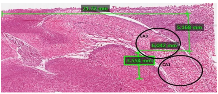
According to the previous work by Holm, the formula above was determined using the number of cells per slab multiplied by the size of the hippocampus extension. To replicate this work, the ITCN ImageJ software plugin was used to count the number of cells per slab. Next, the ImageJ ruler was used to measure the area of the hippocampus. Holm’s formula was applied to obtain the final number of hippocampal cells per subject per area. Finally, the CA1 and CA3 percentage were calculated applying the same formula on the respective areas (Figure 1).
Results and Discussion:
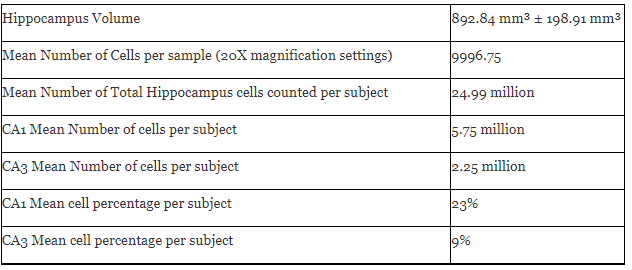
The mean total hippocampus volume was 892.84 mm3 ± 198.91 mm3 using Holm’s methodology. The mean number of cells per sample (20X magnification settings) was 9996.75, using automated ImageJ cell counting. The percentages of hippocampal cells in areas CA1 and CA3 were 9% and 23% respectively, after normalizing by weight. Results are listed in table 1.
The cytoarchitecture of the pig hippocampus is similar to humans. Thus, it is crucial to establish a stereological number of hippocampal cells in pigs as a basis for future studies.
It is important to correct the cell counts in proportion to body weight, as we did, and as Holm did.
Our results showed an average number of hippocampus cells of 24.99 million. This is similar to those reported by Holm (21.61 million).
In this study, the counts of hippocampus cells were divided into two regions of interest: CA1 and CA3. Our results show that the mean number of hippocampus cells observed was 5.75 million and 2.25 million, in the CA1 and CA3 regions respectively. These results are comparable to previous studies. Holm reported 4.12 million cells in the CA1 region and 1.51 million cells in the CA3 region. The results presented here indicate the CA1 and CA3 cell percentages being 23% and 9% respectively, which are similar to the percentages reported by Holm (21% and 12%). It is important to highlight that the proportion of cells is similar in the human hippocampus.
This study provides pig hippocampus data to be used in future studies. So far, only one paper was found to address this topic.[4] However, the use of open source software to stereologically study the hippocampus was not mentioned as part of the reported methodology. The use of open source software (in our case, ImageJ) is a novel and cheaper approach to stereologically analyze the brain tissue.
Limitations: On this study we did not analyze pigs' diets,which might change the hippocampus structure and volume [7] being a variable to be considered in future comparative studies.
Conclusion:
These results corroborate previous findings and demonstrate a novel and cost-effective way to study the hippocampus of pigs in translational neurological research.
Acknowledgements
This study was funded by Lungpacer Medical Inc.
Dear Editorial Team, Clinical Medical Reviews and Reports. My experience with the journal was highly positive. The peer-review process was rigorous, constructive, and completed in a timely manner. The reviewers provided valuable comments that helped improve the quality and clarity of our manuscript. The editorial office was professional, responsive, and supportive throughout all stages of the publication process. Communication was clear and efficient, and any questions were addressed promptly. Overall, I found the journal to maintain high scientific standards and an excellent publication workflow. I would be pleased to consider submitting future work to this journal. Best wishes from, Elena Popa.

It was my pleasure to submit my testimonial concerning the Reviewer Board of our Scientific Journal “Brain and Neurological Disorders”. The Reviewers focused on some modifications and their contribution was helpful. The ladies of our Editorial Office were also supported my efforts. It was my honor to have such a co-operation and I am looking forward for more collaboration.

Dear Grace Pierce, Editorial Coordinator of Journal of Clinical Research and Reports, Thank you for the speedy and efficient peer review process. I appreciate the fact that your peer reviewers do not take months to respond like with some other journals. I would also like to thank the editorial office for responding quickly to my questions. It is an excellent journal. I plan to submit more manuscripts in the future. Best wishes from, Robert W. McGee

Dear Grace Pierce, Editorial Coordinator of Journal of Clinical Research and Reports, Working with you and your team on our recent publication in JCRR has been a truly wonderful and enjoyable experience. The responses were prompt, and the reviewers were patient, constructive, and highly professional. One reviewer in particular gave me the feeling that a professor was carefully reading and commenting on my coursework, which was deeply touching. The entire process was straightforward and hassle‑free, with no tedious online forms to complete. I highly recommend this journal. Best wishes from, DR Aibing Rao, Head of R&D

I Appreciate the Opportunity to Share my Experience with the Journal of Clinical Research and Reports. The peer review process was timely and constructive, and the feedback provided helped improve the quality of our manuscript. The editorial office was professional, responsive, and supportive throughout the process, ensuring smooth communication and efficient handling of the submission. Overall, it was a positive experience collaborating with your team.

Dear Mercy Grace, Editorial Coordinator of Obstetrics Gynecology and Reproductive Sciences, We would like to express our gratitude for your help at all stages of publishing and editing the article. The editors of the magazine answer all the necessary questions and help at every stage. We will definitely continue to cooperate and publish other works in the Obstetrics Gynecology and Reproductive Sciences! Best wishes from, Alla Konstantinovna Politova,
