

Review Article | DOI: https://doi.org/10.31579/2693-2156/165
President of all nations,Morning star hospital, Marthandam, Kanyakumari District, Tamil nadu state, India.
*Corresponding Author: Ramachandran Muthiah, President of all nations,Morning star hospital, Marthandam, Kanyakumari District, Tamil nadu state, India.
Citation: Ramachandran Muthiah, (2026), Acute Rheumatic Fever with Malarial Infection- An Immune Mediated Pathogenesis, J Thoracic Disease and Cardiothoracic Surgery, 7(2); DOI:10.31579/2693-2156/165
Copyright: © 2026, Ramachandran Muthiah. This is an open access article distributed under the Creative Commons Attribution License, which permits unrestricted use, distribution, and reproduction in any medium, provided the original work is properly cited.
Received: 09 April 2026 | Accepted: 20 April 2026 | Published: 11 May 2026
Keywords: febrile episodes; acute rheumatic fever; aso titer; malarial infection; carditis, immune response; duffy blood group antigen
The coexistence of acute rheumatic fever (ARF) with malarial infection is uncommon, its pathogenesis remains unexplained and may related to altered immune status, a unique dual challenge in immunology. Both malaria and ARF can present with high fevers, joint pain (arthralgia), making the clinical diagnosis in endemic areas very difficult. Fever pattern is helpful to diagnose the associated infection with ARF and malaria produces a relapsing fever at 48 hrs interval in plasmodium vivax infection. In ARF, virulence factors, infectious burden and genetic susceptibility play a role. The concept of ‘rheumatogenic’ strains is inadequate and the infectious burden of Strep A (streptococcus A) that “primes” the immune system for dysregulation and self-reactivity during the window period between the onset of Strep A infections and the development of ARF, potentially exacerbating the systemic inflammation and autoimmune responses. The circulating antistreptococcal cross-reactive antibodies and malaria-induced immune complexes aggravate the inflammation of carditis and valvulitis in ARF.
Acute Rheumatic Fever remains a challenge to physicians in developing countries since cyclic fevers with chills synchronous with periodicity may develop in partially immune patients who were previously infected with malarial parasites [1] due to the rupture of infected red blood cells [2]. In those people who were not infected with malarial parasites earlier, the febrile paroxysms are asynchronous and no periodicity since they are not immune to the infection. Altered immune status occurred due to previous infection with P. vivax since it is partially immune as evidenced by 48 hrs periodicity of fever pattern. Similarly repeated pharyngeal infection with group A β hemolytic streptococci as evidenced by raised ASO (anti streptolysin O) titers is necessary to prime the immune response [3] before the first episode of acute rheumatic fever to occur. In tropical countries, there is high prevalence of mixed infections and so, the possibilities of other infections must be evaluated if fever is not responding to routine therapy in acute rheumatic fever.
Etiopathogenesis
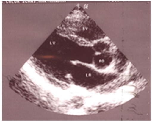
Figure 1: shows Thickened mitral valve tips suggesting Rheumatic involvement in a 12-year-old boy
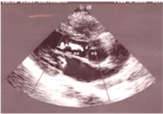
Figure 2: shows mild mitral regurgitation and moderate aortic regurgitation suggesting Rheumatic carditis in a 12-year-old boy
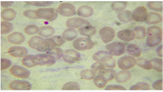
Figure 3: shows Trophozoites of P. vivax in a 12-year-old boy
Malarial fever
Malaria is transmitted through the bite of a female Anopheles mosquito, sporozoites are released from the mosquito salivary glands into host’s skin (dermis), enter the bloodstream, circulate to the liver and invade hepatocytes and differentiate. During this pre-erythrocytic stage as shown in Figure 4, the infection is not apparent clinically, antibodies (Abs) developed against the circumsporozoite surface protein which trap the sporozoites in the skin and reduce their motility to liver cells. Similarly, the Ab-dependent cellular cytotoxicity, interferon-g -producing CD4+ and CD8+ T cells, NK cells and gd T cells destroy the infected liver cells, but the naturally occurring acquired immunity at this stage is not much effective to prevent the recurrent infections in endemic areas. The circumsporozoite protein forms the basis of malaria vaccine and both Abs and CD4+ T cells contribute to 50?ficacy [4].
The infected liver cell will rupture, releasing the merozoites which invade the red blood cells (RBCs) and maturing from rings to trophozoites to form the schizonts. Eventually, the schizonts rupture to release more merozoites into the bloodstream and clinical symptoms of malaria (fever, headache, nausea) occurs during this erythrocytic or blood stage of infection, at this stage, the pro-inflammatory cytokines (IL-1b, IL-6, IFNg, TNFa and IL-12), which enhances phagocytosis and killing of iRBCs (infected red blood cells) by macrophages. However, over-production of these cytokines contributes to malarial pathology and true relapses originate from the dormant liver stages called hypnozoites, which are produced by P. vivax and P. ovale [5].
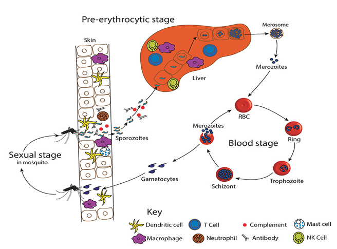
Figure 4: showing the malaria life cycle
According to the ecological assumptions about the thermal physiology of insects, it is found that malaria transmission does not occur at temperature below 16◦C or above 33◦C and at altitude > 2000 m above sea level [4] since the development in the mosquito (sporogony) cannot occur. Definitive diagnosis of malaria generally requires direct observation of malaria parasites in Giemsa-stained thick and thin blood smears. Thick blood smears are more difficult to interpret and the thin smears, in which the parasites are seen within the erythrocytes as in Figure 3, used to determine the species of the infecting parasite [6].
Acute Rheumatic Fever (ARF)
Acute rheumatic fever (ARF) is an immune disorder due to repeated infections (infectious burden) with group A Streptococcus (GAS) [7] and recurrent episodes can lead to rheumatic heart disease (RHD) [8] as shown in Figure 5. The virulence factors as in Figure 6 that are surface-bound (Hyaluronic acid capsules, S protein, Deoxyribonucleases (DNases), streptokinase, immunoglobulin degrading enzyme) well as secreted (streptococcal pyrogenic exotoxin B (Spe B), streptococcal lysozyme streptolysins S and O (SLS and SLO), NAD glycohydrolase (NADase), Hyaluronidase, C5a peptidase [9] induces the colonization, evasion of host immune response and the progress of inflammation and autoimmune reactions.
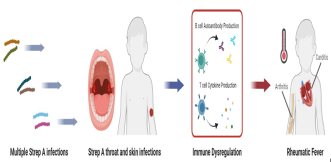
Figure 5: showing the infectious burden of streptococcus A leading to Rheumatic fever
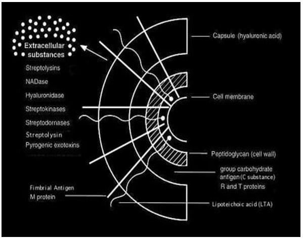
Figure 6: showing the Cell surface structure of Streptococcus pyogenes and secreted products involved in virulence.
Glycoprotein biology (Molecular mimicry)
Molecular mimicry refers to the sharing of antibodies or T-cell epitopes between the host and a microbe. The immune response against streptococcal antigens can lead cross-recognition of heart tissue proteins resulting in rheumatic heart disease (RHD). The rheumatogenic streptococci contain multiple antigenic determinants that partially mimic normal human tissue antigens. These antigens are recognized as foreign by the susceptible host and induces a hyperactive cellular and humoral immune responses. One type involves the recognition of similar structures by antibodies. The alpha-helical structures of protein are found in the N-acetyl-beta-D-glucosamine (S. pyogenes carbohydrate antigen) cross react with antibodies to the heart valve tissue [10]. Another type of molecular mimicry may contribute to immunological cross-reactivity as shown in Figure 7 between diverse molecules, such as DNA and proteins or carbohydrates and peptides [11]. Antibodies induced by group A carbohydrate epitope GlcNAc react with human cardiac proteins, including myosin and laminin, and are capable of causing humoral immune damage. Myosin is an intracellular antigen that is not expressed in human heart valves. M protein shares the epitopes of cross-reaction antibodies with myosin. The streptococcal M protein homology with contractile (myosin, tropomyosin) and interstitial [keratin, laminin, vimentin) proteins and the interstitial reaction of anti-M protein antibody induces the myocarditis whereas the cellular immunity to the shared antigenic determinants contributes the pathogenesis of valvulitis.
Superantigens
Two streptococcal antigens the pepsin generated fraction of M protein and a streptococcal pyrogenic exotoxin (Spe’s) are believed to behave as “Superantigens”. The M protein epitopes not only can trigger the heart cross reactive antibodies, but can also act as superantigens to result in a more widespread immune response by overriding the histocompatibility barrier [12]. These antigens do not bind to antigen binding clefts in T or B cells and can amplify an immune response by clonal expansion of T and B cells, release several cytokines (TNFα, IL I β, IL-6), inducible nitric oxide synthase and adhesion molecules, which could help to localize immune responses to certain tissues. Super antigenic stimulation may help T cells respond to antigens like M proteins suggest a potential mechanism mediating the unrestrained immunologic assault postulated to cause acute rheumatic fever [13].
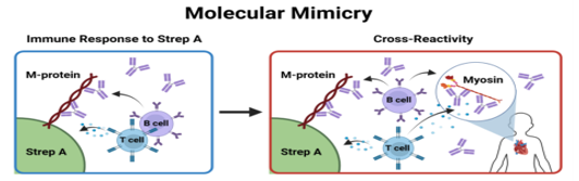
Figure 7: showing the immunological cross reactivity
Rheumatic carditis
The genesis of carditis based on humoral theory due to myocardial damage is uncertain since myocyte necrosis is not a prominent feature [14] and pathological damage is seen in heart valves while no residual damage is observed in myocardium or pericardium after the resolution of ARF.
M protein is the major virulence determinant for S. pyogenese, an alpha-helical coiled coil molecule with extensive variation in structure between Strep A strains, which appears like the fuzz on a tennis ball when viewed by transmission electron microscopy as shown in Figure 8.
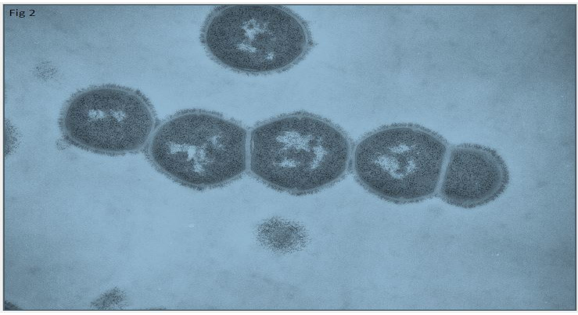
Figure 8: showing the thin section electron micrograph of a chain of streptococci showing the surface M protein. Magnification 50,000x [15]
The N-terminal includes the region coded by the emm gene used for typing Strep A strain types. The C-terminal is anchored in the cell wall, with various repeats (A, B, C, and D) intervening and increasing in sequence conservation between strains. Variation in this structure is known as emm pattern and are designated as patterns A-C (pharyngeal tropism), D (skin tropism), and E (pharyngeal and skin tropism) as shown in Figures 9 and 10.
Collagen has been suggested as the primary target of inflammation in ARF [16], with M-protein binding to type IV collagen in the basement membrane, possibly by the peptide associated with rheumatic fever (PARF) motif of M protein, resulting in autoantibodies to collagen epitopes exposed by M-protein binding (cryptic epitopes as shown in Figure 11) that don’t cross react to M-protein to initiate autoreactivity
[17]. It has been also pointed out that antibodies cross-reactive with myosin were originally identified binding to the valvular endothelium, presumably due to similarity in structure between laminin, myosin, and M-protein [18]. Vascular cell adhesion molecule 1(VCAM 1) may be the connection between humoral and cellular immunity at the surface of the valve. M1 macrophages and the very late antigen 4 (VLA4)/vascular cell adhesion molecule-1 (VCAM-1) pathway may be involved in the inflammatory infiltration and valvular fibrosis in RHD. VCAM 1 gets upregulated at the surface of the endothelium because of the cross-reactive antibodies binding which results in the adherence of CD4+ T cells to the endothelium thereby subsequently infiltrating those cells to the valve [19].
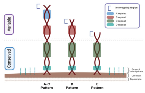
Figure 9: showing the M-protein structure and pattern variety.
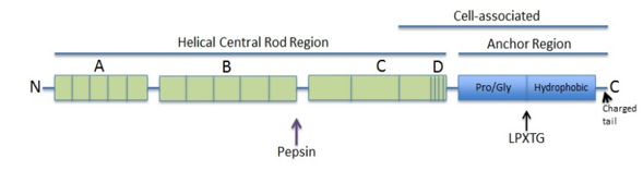
Figure 10: showing the Characteristics of the complete M6 protein sequence
Recent study from Uganda, identified a group of febrile children with echo identified valvular involvement as the sole manifestation, termed as “silent ARF.” [20]. The morphological changes associated with RHD often develop at a later stage of disease and thus are not required for the diagnosis of acute carditis in the setting of ARF. “Latent RHD” can occur without a preceding pharyngitis or ARF and it is more common in India [21]. Some patients with acute rheumatic fever have echocardiographic evidence of thickened mitral leaflet tips as in Figure 1 with mitral and aortic regurgitations as in Figure 2 suggesting the rheumatic carditis as stage B category [22].
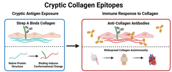
Figure 11: showing the cryptic collagen epitopes [23]
Disruption of immune tolerance
There is no molecular mimicry between the Scl (streptococcal collagen-like) proteins of streptococcus and human collagen [24] and a sustained disparity between Th17 cells and regulatory T cells (Tregs) may disrupt immune tolerance to enhance the susceptibility of the host to streptococcal infection [25]. GAS infection induces the production of TGF-β1, that facilitates the differentiation of Tregs and Th17 cells. Nasopharyngeal-associated lymphoid tissue (NALT) stimulate the antigen uptake, the microfold (M) cells in the NALT overlying epithelium actively sample airborne antigens to activate the mucosal immune response [26]. Prothymosin Alpha induces the pathological immune response of CD8+ T-cells regulated by estradiol receptor alpha (ERα), and also to recognise the type I collagen, in rheumatic heart disease [27].
Epigenetic priming
During the early stage, streptococcus infection activates immunoglobulin G2 autoantibodies (Ig G2), directed against the GlcNAc epitope in the heart valve tissues and these antibodies serve as biomarkers to predict the risk of autoimmune responses following GAS infections [28]. With recurrent infections, the accumulation of IgG2 antibodies may activate the inflammatory response in the valve endothelium and stimulate the inflammatory T-cell subsets such as Th1 and Th17 to cause pro-fibrotic gene expression in endothelial and interstitial cells of cardiac valves [29]. The progression of ARF to RHD, involving epigenetic modification following Strep A mucosal infection and this epigenetic priming along with haemodynamic stress due to transvalvular pressure gradients, initiate valvulitis during ARF through TGF-β1 signalling [30], resulting in progression to chronic RHD characterised by ongoing valvular fibrosis and calcification. Thus, a breakdown of immune tolerance in the host, followed by systemic immune dysregulation, ultimately favouring a pro-inflammatory response to RHD [31].
Immunological periscope
Mucosa-associated invariant T (MAIT) cells recognize MR1 antigens produced during microbial riboflavin biosynthesis and eliciting protective innate immune responses against microorganisms that generate such metabolites. In GAS infection, high activation of human MAIT cells to contribute the initial cytokine response [32] in the pathogenesis of ARF [33]. MR1(Major Histocompatibility Complex class I-related 1) is an immune protein that acts as an "immunological periscope" to detect intracellular infections and it works by presenting small organic molecules (specifically vitamin B metabolites) to MAIT (Mucosal-Associated Invariant T) cells, acting as a rapid sensor for microbes.
Cytokine and chemokine dysregulation
IL-1β-GM-CSF axis NLRP3 Inflammasome (NOD-, LRR- and pyrin domain-containing protein 3) is an intracellular sensor that detects a broad range of microbial motifs) via the induction of pro-inflammatory cytokines, such as IL-1β and IL-18, which in turn stimulate CD4+ T lymphocytes [34], which play a role in the pathogenesis of myocarditis by stimulating Th cells [35] and it is the main source of GM-CSF. The increased production of nterleukin-1β in peripheral blood mononuclear cells in ARF can lead to dysregulated cytokine signaling, specifically the IL-1β-GM-CSF axis (interleukin-1β-granulocyte-macrophage colony-stimulating factor IL-1β-GM-CSF) [36], leading to the specialized migration of TH1 cells to the mitral valve and immunomodulatory drugs [37] are aimed at treating patients at high risk for ARF.
Chemokines
The chemokines, CXCL9/Mig (also known as MIG, or Monokine Induced by Gamma Interferon) is an inflammatory cytokine that belongs to the CXC chemokine family, acts as a chemical signal to recruit immune cells, particularly activated T cells and Natural Killer (NK) cells to sites of inflammation and can enhance T-cell recruitment in valvular tissue lesions [38] and C-C chemokine receptor type 2-positive (CCR2+) macrophages, a specific subset of immune cells primarily derived from circulating blood monocytes. They are actively recruited to sites of inflammation and tissue injury by the chemokine ligand CCL2 (also known as MCP-1) in autoimmune diseases and tissue fibrosis, and suggest that they are critical for valve remodeling in RHD [39] as shown in Figure 12.
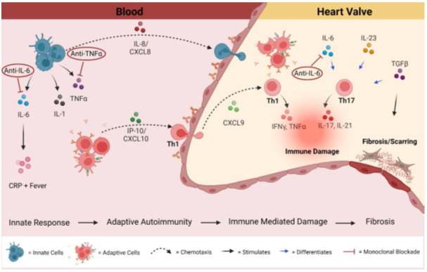
Figure 12: showing the Cytokine and chemokine mediated autoimmunity in blood and heart valve tissue during acute rheumatic fever and rheumatic heart disease.
Genetic correlations
HLA genotypes
HLA antigen may account for individual susceptibility to rheumatic fever and only a few develop RHD, and the incidence is three times higher in monozygotic than in dizygotic twins [40]. There is a significant association between specific HLA class II antigen, HLA DR2 in blacks, HLA DR4 in whites, HLA DR4 in the United States and Saudi Arabia, HLA DR3 and DQw2 in India, HLA-DR15 and DRB5 alleles with mitral valve disease (10%) and rheumatic fever [41]. In contrast, Ozkan et al study revealed HLA-DR5 is a protective factor against RHD [42] and B cell alloantigen D8/17 have shown a strong susceptibility to rheumatic fever [43]. The distribution of class I HLA antigens in rheumatic fever is inconclusive.
Duffy Antigen Receptor
HLA BW53 protects against malaria due to a polymorphism in TNFα promoter region and in Plasmodium vivax, that determines resistance is the genetic polymorphism of parasite receptor known as Duffy Antigen Receptor for Chemokines (DARC) [44], [45] as shown in Figure 13. An approach to identify small molecule inhibitors of DARC should prove to be of benefit in drastically reducing P. vivax malaria. Duffy-negative allele, lack of expression of chemokines receptor and Chemokines sink to amplify the immune response that does not allow the entry of the parasite to the red blood cells (RBCs) [46].
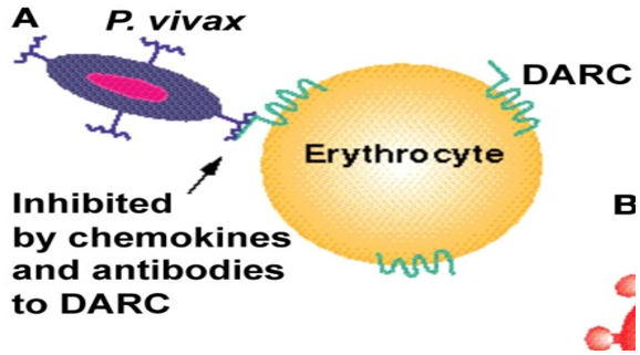
Figure 13: A) The human chemokine binding protein DARC is expressed on erythrocytes and is used by P. vivax to gain entry to the cell.
It can be inhibited by a monoclonal antibody to DARC, Fy6, and by chemokines such as CXCL1 and CXCL8 [47]
There are two kinds of alleles related to the receptors DARC: Fya and Fyb, which identifies four possible phenotypic presentations: homozygous Fy (ab−) (absence of the receiver or null), homozygous Fy (a+ b+) and heterozygous Fy (a− b+) and Fy (a+ b−). These receptors belong to the family of seven transmembrane molecules, initially recognized as a receptor for the Plasmodium vivax in human red blood cells. They are recognized as a “promiscuous” receptor which is able to bind both CC (β-chemokines, primarily attract monocytes, macrophages, and T lymphocytes to the sites of chronic inflammation) and CXC chemokines (α-chemokines, predominantly recruit neutrophils to sites of acute inflammation) and to have a cleaner role of these molecules [48].
Mannose-binding lectin (MBL)
In RHD patients, there is significantly increased levels of mannose-binding lectin (MBL- an innate mediator) occurs. MBL acts as a primary pathogen-recognition molecule that drives the lectin complement pathway and this cascade exacerbates inflammation and tissue damage, targets the body’s own heart valves, causing severe valvular damage. There is a beneficial role for MBL deficiency in these patients [49].
‘Vitamin C ‘hypothesis
The genus Homo lost the ability to synthesize Vitamin C corresponding with the evolution of color vision [50] and Streptococcus got adapted to infect species that have a limited ability to synthesize vitamin C, such as humans and guinea pigs [51]. Humans, in contrast to many animals, are incapable of synthesising this vitamin (Vitamin means that one has to eat it) and are totally dependent on its dietary intake. Szent-Györgyi, in October 1937, was awarded the Nobel Prize for Physiology or Medicine “for his discoveries in connection with the biological combustion processes, with special reference to vitamin C (antiscorbutic factor from lemon juice and named it as vitamin C and Szent-Györgyi had suspected that hexuronic acid was, in fact, vitamin C)”. The concept of mega doses of vitamin C have a beneficial effect and Linus Pauling, winner of two Nobel prizes (Chemistry Prize in 1954, Peace Prize in 1962) suggested that the intake of vitamin C should be about 2.3 g per day for the maintenance of general health, and increased intake up to 10 g/day for combating infectious diseases, including the common cold as well as prevention and cure of cardiovascular diseases and cancer. In humans, a regular intake of about 2 g vitamin C/day should protect a moderate smoker (10–15 cigarettes/day) from emphysema [52].
Immune signature (Immune Priming)
There is marked heterogeneity in the ARF autoantibody profile [53]. Both serological and epidemiological data suggest that repeated GAS infections prime the immune system and break the immune tolerance to develop ARF [54],[55]. There is increasing evidence that GAS skin infections might also be a trigger for ARF, either directly or as part of immune priming [56]. The repeated exposures of GAS drive the exaggerated IgG3 responses to M-proteins observed in ARF leads to immune dysregulation [57] as in Figure 14.
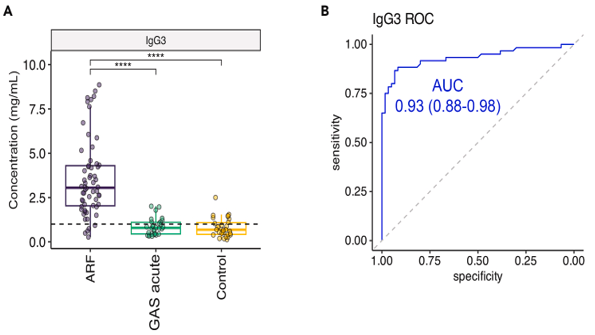
Figure 14: showing the elevated IgG3 in ARF ((A) illustrating the distribution of IgG3 concentrations across study groups. (B) A receiver operator curve (ROC) curve comparing ARF to all controls [58].
The observation of elevated IL-6, complement C4 and CRP (C4-CRP-IL6 inflammatory triad), points clearly to the inflammation underlying ARF [59] and the immune signature is shown in FIgure 15.
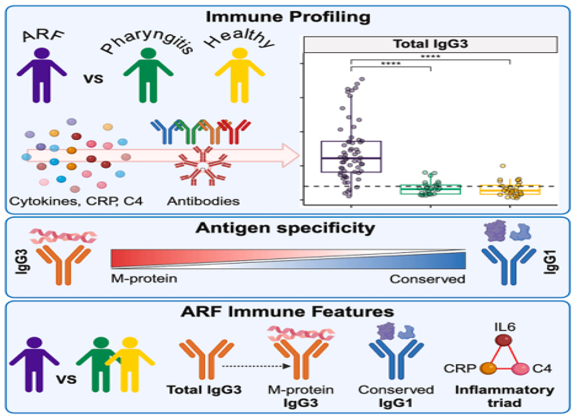
Figure 15: showing the immune signature comprising inflammatory markers, IgG3, and Streptococcus pyogenes-specific antibodies in acute rheumatic fever.
Epidemiological surveys
In tropical countries, mixed infections of streptococci both in the throat and skin due to various strains are more common. Group C or G streptococcus are frequently isolated from the throat than Group A streptococcus in Rheumatic Fever patients in high-risk indigenous populations of Aboriginal communities in Australia [60], [61] and Māori and Pacific Peoples in Aotearoa New Zealand [62] and they can occur virulence factors by horizontal transmission from Group A streptococci. There is a high prevalence of rheumatic fever in patients with skin lesions even though streptococcal pyoderma does not cause Rheumatic fever [63] since streptolysin O is irreversibly inactivated by lipids (probably cholesterol) present in the skin and unable to induce increased magnitude of the immune response to produce rheumatic fever [64]. Earlier studies categorically differentiated the nephritogenic and rheumatogenic strains, but recent studies displace this concept by suggesting that both mucosal and skin infection with GAS can lead to long-term cardiac sequelae [65]. Epidemiological surveys are necessary to find out whether any higher incidence of acute rheumatic fever in endemic areas of malaria and any association with specific blood group antigen especially with duffy factor. More recent progress to diagnose GAS pharyngitis include Nucleic Acid Amplification Tests (NAATs), which have much better sensitivity and specificity than RADTs (rapid antigen detection test) [66]. The difference between GAS pharyngitis and viral pharyngitis is shown in Table 1
| S.No | Streptococcal pharyngitis | Viral pharyngitis |
1.
2.
3
4
5. | headache, abdominal pain, nausea, vomiting and no diarrhea.
Fever of more than 38◦C and lack of cough.
Tendor or swollen anterior cervical nodes
tonsillar exudates (white patches/pus on the tonsils) or swelling
Pain with swallowing | Prominent catarrhal, symptoms common cold-like symptoms (watery eyes, running nose) Fever with dry cough may occur
Cervical nodes not enlarged
Tonsils and throat are red with vesicles and ulcers
Uncomfortable throat |
Table 1: showing the difference between streptococcal pharyngitis and viral pharyngitis
Therapeutics
ARF Treatment requires immediate antibiotic therapy to eradicate the streptococcus bacteria, anti-inflammatory medications (like aspirin) to control joint and heart inflammation, and long-term secondary antibiotic prophylaxis {SAP) to prevent recurrences. ASA (Aspirin (Acetylsalicylic Acid) (60 to 100 mg/kg/day in divided doses until symptom resolution) is the most commonly used NSAID in the treatment of ARF with a dramatic response and resolution of fever and arthritis in 1 to 3 days. However, there is no documented evidence showing superior efficacy of any drug (steroids or NSAIDs) in reducing the risk of rheumatic valve damage (RVD) in patients with ARF [67]. The recent meta-analysis of 51 studies on secondary prophylaxis showed that good adherence to reduce the progression of the disease by 71% [68].
GAS is exclusively a human pathogen, co-evolved with Homo Sapiens and exists in a carrier state in the throat and skin in about 20% of children [69]. The antibiotic, cephalosporins have some superiority in eradicating GAS infections since they reduce the penicillinase-generating pharyngeal flora, which are implicated in GAS carrier state [70]. Recently it is found that Immunoglobulins are of no value in the management of acute rheumatic fever [71].
In 1965, American chemist Robert Burns Woodward was awarded the Nobel Prize in Chemistry for the first total synthesis of quinine, one of the earliest and most effective antimalarial drugs. Malaria treatment via standard antimalarial medications (e.g., Artemisinin-based Combination Therapy or chloroquine), depending on the specific parasite strain (P. falciparum or P. vivax) and local resistance patterns is widely practiced. Mefloquine is not recommended for patients with cardiac conduction defects or who are taking β blockers. Arrhythmogenic effects of chloroquine are not seen at normal doses, but do occur with rapid intravenous infusion or massive overdoses. In areas co-endemic for P vivax and Plasmodium falciparum, there is an increased risk of P. vivax parasitaemia with sexual stages [72] and it is a rationale for opportunistically eradicating P. vivax hypnozoites from the liver, an approach termed universal radical cure. Most endemic countries currently recommend a low-dose regimen of primaquine (total dose 3·5 mg/kg) for the radical cure of P vivax malaria, administered over 14 days [73],[74]. This prolonged treatment limits the daily dose of primaquine to 0·25 mg/kg per day to improve tolerability and to reduce the risk of drug-induced haemolysis [75].
Immunomodulatory therapy
There is a complete lack of therapies that can halt the progression of ARF to RHD [76]. The immune profile suggests that IL-6 or TNFα blockade could be effective for suppression of this acute disease state.
Il-6 blockade
IL-6 blockade by the humanized anti-IL-6R IgG1 monoclonal antibody, the Tocilizumab (TCZ) has proven effective in multiple autoimmune diseases. Elevated circulating levels of IL-6 in ARF and RHD suggests that TCZ is a promising immunomodulating therapy. According to genetic studies, including a New Zealand study, it is found that IL-6 related variants increase the susceptibility to RHD [77]. The inflammatory parameters, CRP decrease and reduced immune cell infiltration by TCZ are beneficial in ARF and it also modulates M2 macrophages known to have a pathological role in fibrosis of valves in RHD [78]. TCZ has been proposed as a promising broad action immunomodulating drug for a large variety of “chronic intractable immune mediated diseases,”. Tocilizumab has also shown to reduce serum IgG3 levels clinically and the available evidence suggests that ARF and RHD should be included under this therapy [79], [80].
TNFα Blockade
TNFα is variably increased in Acute phase of RF (Rheumatic fever) [81], shown to regulate pro-inflammatory cytokines including IL-1, IL-6, GM-CSF, and IL-8/CXCL8 and TNFα blockade in clinical trials to dampen this pro-inflammatory “cascade” with its ability to reduce the cell mediated tissue ingress and systemic autoimmunity, it is worth exploring as a therapeutic option for ARF and RHD.
TGFβ inhibition
TGFβ is a pleiotropic cytokine growth factor, and is known to cause cardiac damage. Increased TGFβ has been observed in RHD heart valves, with significant correlations to immune cell infiltration, neovascularization, calcification, myofibroblast proliferation, and valvular fibrosis [82]. Inhibition of TGFβ signaling will make an understanding of the pathogenesis RHD, and to evaluate its value as a target for intervention.
IP-10/CXCL10 inhibition
The increased levels of IP-10/CXCL10 seen in ARF and its association with adverse outcomes in post-surgery in RHD, warrants further investigation to ameliorate the disease.
Rituximab
It is a chimeric anti-CD20 monoclonal antibody therapy that acts via transient B cell depletion. Since antibody deposition on heart valves is important to the pathogenesis of ARF/RHD, B cell depletion may be a possible therapeutic approach to control the disease [83].
Hydroxychloroquine
Hydroxychloroquine (HCQ) suppresses an interleukin-1β-granulocyte-macrophage colony-stimulating factor cytokine axis, reported to be dysregulated in peripheral blood mononuclear cells of acute rheumatic fever. HCQ treatment was associated with control of inflammatory markers, pericarditis and stabilization of progressive carditis [84].
Strep A vaccine
The hypothesis of immune priming suggests that multiple infectious events are required to cause ARF and a precise extent of strain coverage is needed to prevent ARF by vaccination, but whether long term immune memory generated to Strep A is uncertain [85],[86].
Acute rheumatic fever (ARF) is an autoimmune response to untreated Group A streptococcal throat infections, whereas malaria is a parasitic infection. These two distinct conditions are often medically intertwined due to overlapping symptoms such as high fevers and joint pain which can make it difficult to differentiate them in tropical regions of India. Studies have shown diagnostic overlaps where patients (especially children) test positive for malaria but also meet the criteria for ARF. Failing to treat ARF can lead to permanent Rheumatic Heart Disease. Clinicians rely on rapid diagnostic tests for malaria, throat cultures, antistreptolysin O (ASO) titers and echocardiography to distinguish and treat both diseases appropriately. India bears a significant burden of both diseases and treating ARF aggressively alongside malaria is crucial for preventing RHD. Local healthcare infrastructure continues to focus on improving both public awareness and early echocardiographic screening.
Dear Editorial Team, Clinical Medical Reviews and Reports. My experience with the journal was highly positive. The peer-review process was rigorous, constructive, and completed in a timely manner. The reviewers provided valuable comments that helped improve the quality and clarity of our manuscript. The editorial office was professional, responsive, and supportive throughout all stages of the publication process. Communication was clear and efficient, and any questions were addressed promptly. Overall, I found the journal to maintain high scientific standards and an excellent publication workflow. I would be pleased to consider submitting future work to this journal. Best wishes from, Elena Popa.

It was my pleasure to submit my testimonial concerning the Reviewer Board of our Scientific Journal “Brain and Neurological Disorders”. The Reviewers focused on some modifications and their contribution was helpful. The ladies of our Editorial Office were also supported my efforts. It was my honor to have such a co-operation and I am looking forward for more collaboration.

Dear Grace Pierce, Editorial Coordinator of Journal of Clinical Research and Reports, Thank you for the speedy and efficient peer review process. I appreciate the fact that your peer reviewers do not take months to respond like with some other journals. I would also like to thank the editorial office for responding quickly to my questions. It is an excellent journal. I plan to submit more manuscripts in the future. Best wishes from, Robert W. McGee

Dear Grace Pierce, Editorial Coordinator of Journal of Clinical Research and Reports, Working with you and your team on our recent publication in JCRR has been a truly wonderful and enjoyable experience. The responses were prompt, and the reviewers were patient, constructive, and highly professional. One reviewer in particular gave me the feeling that a professor was carefully reading and commenting on my coursework, which was deeply touching. The entire process was straightforward and hassle‑free, with no tedious online forms to complete. I highly recommend this journal. Best wishes from, DR Aibing Rao, Head of R&D

I Appreciate the Opportunity to Share my Experience with the Journal of Clinical Research and Reports. The peer review process was timely and constructive, and the feedback provided helped improve the quality of our manuscript. The editorial office was professional, responsive, and supportive throughout the process, ensuring smooth communication and efficient handling of the submission. Overall, it was a positive experience collaborating with your team.

Dear Mercy Grace, Editorial Coordinator of Obstetrics Gynecology and Reproductive Sciences, We would like to express our gratitude for your help at all stages of publishing and editing the article. The editors of the magazine answer all the necessary questions and help at every stage. We will definitely continue to cooperate and publish other works in the Obstetrics Gynecology and Reproductive Sciences! Best wishes from, Alla Konstantinovna Politova,
