
Case Report | DOI: https://doi.org/10.31579/2834-5142/015
1 Renal department, Mid and South Essex University hospitals; Basildon & Thurrock University Hospital; UK
*Corresponding Author: Michael Fawzy, Clinical lead and Renal consultant, Renal department, Basildon & Thurrock University Hospital
Citation: Nikita B Jolapara, Prabhvir Marway, Michael Fawzy (2022). Acute Medical (bedside) Peritoneal Dialysis Catheter insertion in Intensive Therapy Unit Setting, an Overlooked Renal Replacement Therapy option, A Case report Study. International Journal of Clinical Nephrology. 4(1); DOI:10.31579/2834-5142/015
Copyright: © 2022 Michael Fawzy. This is an open-access article distributed under the terms of the Creative Commons Attribution License, which permits unrestricted use, distribution, and reproduction in any medium, provided the original author and source are credited.
Received: 06 December 2021 | Accepted: 24 December 2021 | Published: 03 January 2022
Keywords: medical/bedside peritoneal dialysis catheter insertion; acute kidney injury (aki); intensive therapy/care unit (itu); covid-19; peritoneal dialysis; haemofiltration
A 56-year-old woman with a background of long-standing type II diabetes and chronic kidney disease presented to hospital with fevers, cough and shortness of breath. She had been self-isolating due to COVID-19 a week prior to admission. Her admission resulted in a long stay in intensive care unit with acute kidney injury. Due to her hypercoagulability state, multiple attempts of haemodialysis and filtration failed resulting in the first attempt of bedside (non-fluoroscopic guided) insertion of peritoneal dialysis catheter in an ITU setting in the region of Essex. We note that usually in an ITU setting, haemodialysis is commonly used. Our case therefore serves as an important reminder that peritoneal dialysis (PD) can serve a significant value for patients with acute kidney injury when haemodialysis cannot help in.
Intensive Therapy units (ITUs) are known to support hemofiltration in an acutely unwell patient. This was extremely useful especially in patients who presented with COVID-19 and acute kidney injury due to severe COVID sepsis. It is important to recognise that there is potential to extend this service and provide Acute peritoneal dialysis when haemofilteration can’t be safely maintained due to a variety of factors (access problems, hemodymamic instability, logistics due to unavailable haemofilters etc) (13).
Peritoneal dialysis (PD) is usually a procedure performed by surgeons and undertaken in a surgical setting. Post insertion there is a minimum of 2-4 weeks wound healing time before starting PD fluid exchange. Now days, the procedure can be performed in a procedure room by a nephrologist via fluoroscopy. This can be very useful due to:
a- It does not require the involvement of a surgical team so it can bypass surgical list backlog.
b-It can be done for those with high risk to general anaesthesia (eg; poor cardiac function, advanced COPD etc).
c- Another added bonus of bedside insertion is that peritoneal dialysis can be started earlier without a waiting period compared to surgical PD catheter due to less trauma elicited.
Our case today was done bedside (blind Seldinger technique without fluoroscopic guidance) [4]
Our case is a 56-year-old female, a care home nurse, who presented with a week history of fevers, cough and shortness of breath. Her symptoms persisted and as per National Health Service (NHS) guidance she began self-isolation. She presented to the hospital after her symptoms progressed and found it difficult to breath with admission oxygen saturations of 64% on room air. A nasopharyngeal swab for SARS-CoV-2 PCR was positive on admission. Her background medical history included hypertension, Type 2 diabetes mellitus, and chronic kidney disease since 2017 with a baseline of creatinine 142umol/L.
On presentation to the emergency department, the patient was noted to be in clear distress. Her respiratory rate was 28 breaths per minute, oxygen saturations 96% on 15L of oxygen provided, blood pressure of 164/73 mmHg, heart rate of 92 beats per minute and temperature of 38.5C. On clinical examination her chest was clear on auscultation bilaterally.
Her admission arterial blood gas on 15L O2 showed Type 1 respiratory failure with pH 7.38, PO2 8.8, PCO2 4.6, HCO 21.4, BE -4.1, Lac 0.5 Glu 9.0. Her biochemistry showed lymphopenia of 0.68 10*9/L, sodium of 133mmol/L, potassium 4.3mmol/L, urea 31.8mmol/L, Creatinine 652umol/L, CRP 417 mg/L.
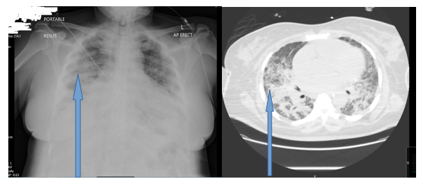
Her chest x-ray was consistent with COVID-19 pneumonia.
Due to her hypoxic ABG, pH 7.21, PO2 5.7, PCO2 5.6, HCO 14.2, BE -12.4, Lac 2.4 and ongoing respiratory distress, she was intubated in resuscitation suite and admitted into ITU for ventilation support.
During her stay in ITU, she started to become anuric with worsening acute kidney injury (AKI) so she was established on continuous veno-venous haemofiltration (CVVH). Unfortunately, she developed bilateral stroke on top of her COVID 19 pneumonia and Multi Drug Resistant pseudomonas infection. The history was so far typical for COVID but sadly to complicate the prognosis, she underwent multiple attempts of Central Venous Catheters (These accesses were needed to maintain CVVH support) as they were clotting within few hours of insertions despite being on continuous infusion of maximum dose of heparin due to her hypercoagulable state. During renal ward round in ITU, He realized that patient was not achieving enough clearance and ultrafiltration due to access problem. This has reflected on inability to wean the patient off high oxygen requirement of ventilator (she was on FiO2 of 100 %). It was obviously a failing CVVH and ITU team was running out of options.
Therefore, it was decided to step in and offered to put medical PD catheter and start PD for her. He examined her abdomen and there was only a scar of previous caesarean section with no obvious hernias.
After calling patient’s next of kin and obtaining the consent based on medical evaluation of patient’s best interest. ITU team was intructed to hold heparin infusion for 2 hours.
After scrubbing, the abdomen is prepped, about 2 cm below the umbilicus a small midline horizontal incision (2-3 cm) was made under local anaesthetic. Blunt dissection of subcutaneous tissue was carried out reaching the anterior rectus sheath. 18g needle was then vertically inserted through the rectus sheath until it entered the abdominal cavity. The needle was then pointed toward the left lower quadrant (LLQ) but not advancing more than another 1-2 cm. Guide wire then is passed through the needle aiming at LLQ. Over the guide wire the trocar was passed through the rectus sheath until it entered the abdominal cavity. This was to dilate the insertion point. The trocar was removed and reinserted this time being inside a peel away sheath. Once the instrument was in the abdominal cavity pointing to the LLQ, the peel away sheath is fed forward with trocar kept fixed. When peel away sheath is well in place, trocar and the guide wire were removed leaving the sheath behind. The 63 cm PD catheter was then passed through the peel away sheath and as the catheter is advanced, the sheath is pulled out and peeled away until all was out and the catheter was in the abdomen up to the proximal cuff. The cuff was pushed down to sit on the rectus sheath. At this time the catheter was tested to make sure that a normal saline flow in with no resistance and also the efflux was clear. From the lateral side of the abdominal incision, more local anaesthetic was given and the catheter was advanced subcutaneously using the tunnelling tool and aiming for the previously marked exit site in the lower part of the abdomen. It is made sure that the distal cuff of the catheter is about 3-5 cm proximal to the exit site and the tunnel is not immediately under the skin. The incision was sutured and dressed with Mepore dressing over the insertion site and the PD catheter exit site, and the PD catheter will be looped and secured with Mefix tape. A titanium and PD Extension set will be fitted and the catheter anchored securely. The tube was anchored to prevent trauma and to keep it immobilized at all times.
The patient started PD within 2 hours of the insertion. She was kept on assisted automated peritoneal dialysis (AAPD) for three weeks with average daily ultrafiltration of 2.5-3 litres. This enabled ITU team to wean her off the ventilator. Later on, patient was stepped down to the renal ward after 43 days in ITU. She carried on PD for another 2 months then her urine output started to improve. Her renal function started to show signs of improvement. After 4 months of daily AAPD, PD was stopped and patient was kept on close follow up with regular blood tests. At that stage, patient was still not verbalizing or able to engage in any conversation due to the double stroke she sustained in ITU.
Later on, the Renal consultant attempted removal of PD catheter bed side under local anaesthetics. I palpated the SC cuff and infiltrated 5ml of Lidocaine 1% around it. A kin incision of 2 cm over the cuff then dissecting the tissues until the first cuff was freed. Then, another 5ml of Lidocaine was infiltrated below the umbilicus. 2 cm Skin incision sub umbilical was done and dissected the SC tissues down to peritoneum to free the inner cuff. Finally, both cuffs were freed and the whole tube was removed successfully.
During the ward admission, her renal function gradually improved, and she was successful weaned off peritoneal dialysis with a new renal baseline creatinine of 212umol/L. Her PD tube was removed at the bedside as detailed above.
The patient underwent a percutaneous endoscopic gastrostomy (PEG) insertion for feeding and nutritional support as a result of her stroke. She further received neuro-rehabilitation therapy in a specialized centre. Miraculously, patient showed progressive improvement and then successfully discharge home. The patient was fully independent and AMT (mini mental capacity test) was 10/10. She had a new renal baseline function and no longer requiring a long-term renal replacement therapy (RRT).
Haemofiltration is commonly used in ITU settings. Our case therefore serves as an important reminder that peritoneal dialysis can serve a significant value for patients. [7]
Patients who presented with severe COVID-19 sepsis and acute kidney injury, with or without a background of chronic kidney disease (CKD), were beneficiaries of haemofiltration in the ITU setting. It is important to recognise that there is potential to extend this service and provide peritoneal dialysis when haemodialysis can’t be safely maintained due to a variety of factors (e.g.; hypercoagulable states or if capacity to provide haemodialysis is limited). [11]
In this case report we present the first case of a medical peritoneal dialysis insertion that successfully attempted in ITU setting in Essex for a patient with severe COVID sepsis and AKI, with a beneficial short- and long-term impact for the patient. It obviously managed to keep dialysis ongoing as short-term benefit and protected the residual renal function as long term benefit.
Acute kidney injury is very common in critically ill patients and ICU patients. Usually, it is quite common to have renal replacement therapy in such setting. 5-6% of AKI on ICU required RRT in a prospective multinational study carried out by Abraham et al. [12] ]This was consistently seen across 23 participating countries. Peritoneal dialysis is often overlooked in an ICU setting with continuous RRT or intermittent haemodialysis favoured more in developed countries. [16,21] PD dialysis has its advantages due to its ease of administration, technicality, low bleeding risk and suitability in cardiovascular instability.[10]
There has been high incident rate of AKI during COVID pandemic and generally COVID patients tend to be in a hypercoagulable state. [17] This is worsened by the fact that patient was in ITU and had background morbidities that tend to substantially augment the hypercoagulable state. With these two factors this can put the patient in a much-compromised state as seen in this case report.
There has been established renal replacement therapy patients with COVID-19 infection into the ITU setting with demonstrations of the prevalence of AKI requiring renal replacement therapy and ITU admission, secondary to COVID-19 infection. [8]
A large retrospective cohort study of 4264 critically ill COVID-19 patients [6], of which 143 were dialysis patients and 521 were CKD patients, indicated poorer outcomes in those with pre-existing kidney disease. Dialysis patients had a shorter time from symptom onset to ICU admission (median days: 4), compared to other groups (median days: 7 for CKD, 7 for those without pre-existing renal disease). 50% of dialysis and CKD patient died within 28 days of ITU admission, compared to 35% in patient s without pre-existing kidney disease (Hazard Ratio (HR) 1.41 for dialysis patients, HR 1.25 for CKD patients). [6]
A small retrospective case series [7] indicated fewer poor outcomes. From 14 patients on HD, or previously advanced CKD and now on HD secondary to COVID-19, four (29%) required mechanical ventilation and continuous RRT. One underwent ECMO for a period of three days. From these 14 patients, two died from ARDS (14%), and nine (64%) were discharged from hospital. Another retrospective case series [5] of 57 HD and 2 PD patients showed that eight (14%) required mechanical ventilation, and of these, three (38%) needed continuous RRT. [5]
The remaining eight papers focused on AKI secondary to COVID-19 infection. A significant minority of patients with COVID-19 develop an AKI, ranging from 3-15% [23]. This AKI is characterised by tubulointerstitial injury without glycosuria [4]. For patients that required ITU admission, this incidence was much higher, ranging from 50-78% [3,8]. This subset of patients with AKI (including AKI on CKD) and COVID -19 infections appear to have an alarmingly high mortality rate, compared to those patients that do not develop an AKI [21]. This may in part be explained by a strain on resources to provide effective RRT via haemodialysis. Some studies indicate the effectiveness of urgent peritoneal dialysis catheter placement as an alternative and reflect the patient case described in this report [8,16].
Six studies (484 participants) compared high volume PD to daily haemodialysis, extended daily haemodialysis, or continuous renal replacement therapy. It was concluded that PD made little or no difference to all-cause mortality or kidney function recovery compared to haemodialysis. It was mentioned that PD might sometimes decrease ultrafiltration compared to other modalities nevertheless, It is uncertain whether PD compared to other modalities has any effects on weekly delivered Kt/V, correction of acidosis. [10]
The authors declare that there is no conflict of interest.
Dear Editorial Team, Clinical Medical Reviews and Reports. My experience with the journal was highly positive. The peer-review process was rigorous, constructive, and completed in a timely manner. The reviewers provided valuable comments that helped improve the quality and clarity of our manuscript. The editorial office was professional, responsive, and supportive throughout all stages of the publication process. Communication was clear and efficient, and any questions were addressed promptly. Overall, I found the journal to maintain high scientific standards and an excellent publication workflow. I would be pleased to consider submitting future work to this journal. Best wishes from, Elena Popa.

It was my pleasure to submit my testimonial concerning the Reviewer Board of our Scientific Journal “Brain and Neurological Disorders”. The Reviewers focused on some modifications and their contribution was helpful. The ladies of our Editorial Office were also supported my efforts. It was my honor to have such a co-operation and I am looking forward for more collaboration.

Dear Grace Pierce, Editorial Coordinator of Journal of Clinical Research and Reports, Thank you for the speedy and efficient peer review process. I appreciate the fact that your peer reviewers do not take months to respond like with some other journals. I would also like to thank the editorial office for responding quickly to my questions. It is an excellent journal. I plan to submit more manuscripts in the future. Best wishes from, Robert W. McGee

Dear Grace Pierce, Editorial Coordinator of Journal of Clinical Research and Reports, Working with you and your team on our recent publication in JCRR has been a truly wonderful and enjoyable experience. The responses were prompt, and the reviewers were patient, constructive, and highly professional. One reviewer in particular gave me the feeling that a professor was carefully reading and commenting on my coursework, which was deeply touching. The entire process was straightforward and hassle‑free, with no tedious online forms to complete. I highly recommend this journal. Best wishes from, DR Aibing Rao, Head of R&D

I Appreciate the Opportunity to Share my Experience with the Journal of Clinical Research and Reports. The peer review process was timely and constructive, and the feedback provided helped improve the quality of our manuscript. The editorial office was professional, responsive, and supportive throughout the process, ensuring smooth communication and efficient handling of the submission. Overall, it was a positive experience collaborating with your team.

Dear Mercy Grace, Editorial Coordinator of Obstetrics Gynecology and Reproductive Sciences, We would like to express our gratitude for your help at all stages of publishing and editing the article. The editors of the magazine answer all the necessary questions and help at every stage. We will definitely continue to cooperate and publish other works in the Obstetrics Gynecology and Reproductive Sciences! Best wishes from, Alla Konstantinovna Politova,
