
Review | DOI: https://doi.org/10.31579/2690-8794/245
1Emergency Medicine Department, Gülnar Goverment Hospital,Mersin, Türkiye
2Hatay Mustafa Kemal University, Faculty of Medicine, Department of Emergency Medicine, Hatay
3Emergency Medicine Department, Arsuz Goverment Hospital,Hatay, Türkiye
*Corresponding Author: : Ali Karakuş, Hatay Mustafa Kemal University, Faculty of Medicine, Department of Emergency Medicine, Hatay
Citation: Pınar Baydar Yücel, Ali Karakuş, Mustafa Polat, Anıl Yoldaş, (2024), Acute Ischaemic Stroke and Thrombolytic Therapy,Clinical Medical Reviews and Reports; 6(10): DOI: 10.31579/2690-8794/245
Copyright: © 2024 Ali Karakuş, this is an open-access article distributed under the terms of the Creative Commons Attribution License, which permits unrestricted use, distribution, and reproduction in any medium, provided the original author and source are credited.
Received: 09 December 2024 | Accepted: 16 December 2024 | Published: 23 December 2024
Keywords: Acute ischaemic stroke; Thrombolytics; Emergency service
Acute ischaemic stroke (AIS) is a preventable and treatable disease with high mortality and morbidity, accounting for 80% of cerebrovascular events. Hypertension (HT), atrial fibrillation (AF) and diabetes mellitus (DM) are risk factors in AIS. The diagnosis is made by starting thrombolytics within the first 4.5 hours after anamnesis, neurological examination and exclusion of haemorrhage by computed tomography.
Acute ischaemic stroke (AIS) is defined as damage to neuronal tissues as a result of decreased blood flow to some or all of the brain tissue. It accounts for 80% of cerebrovascular events. It is a disease with high mortality and morbidity and acute ischaemic stroke is the most common cause of death after coronary artery diseases and cancers in developed countries[1]. Early recognition and rapid treatment is important in reducing mortality and morbidity. Intravenous (IV) recombinant tissue plasminogen activator (rt-PA) is the medical treatment [2].
With this case report, we aimed to increase the awareness of the positive effect of early diagnosis and thrombolytic treatment initiated in the emergency department on the prognosis of the patient and the prompt administration of thrombolytics in emergency departments.
Case
A 50-year-old female patient was brought to the emergency department with a complaint of speech disorder that started about 2 hours ago. She had known congestive heart failure (CHF), AF and HT diseases and was using acetylsalicylic acid and beta blocker drugs. The general condition of the patient was moderate, consciousness was clear, coopere GKS 15 aphasia and right central facial paralysis were present. Other neurological and cisitemic examinations were normal. Blood pressure was 140/80mm/Hg, pulse 130/min, sat 98%, temperature 36C. ECG was compatible with AF. no pathological value was found in blood tests, INR was 1, APTT was 30 seconds, no bleeding area was seen in the brain tomography taken quickly, simultaneous diffusion magnetic resonance (MR) was taken and neurology was consulted. The MRI revealed a diffusion restricting area in the cortex and white matter at the level of the left frontoparietal lobes, hyperintense on diffusion weighted imaging (DAG) and hypointense on image diffusion coefficient (ADC) maps (Figure 1). Alteplase IV (0.9 mg/kg, maximum dose 90 mg, for 60 minutes, initially 10% of this dose was given as a bolus for 1 minute) was started as thrombolytic treatment in the emergency department at approximately the 3rd hour. After the treatment, the patient was admitted to neurology intensive care unit for follow-up. After 24 hours of observation, the patient was taken to the ward and followed up after the absence of complications and clinical improvement. The patient was discharged with healing at the end of the 5th day after clinical improvement, improvement of aphasia, and improvement of central facial paralysis, and neurology outpatient clinic controls showed that the patient recovered without any sequelae after 1 month.
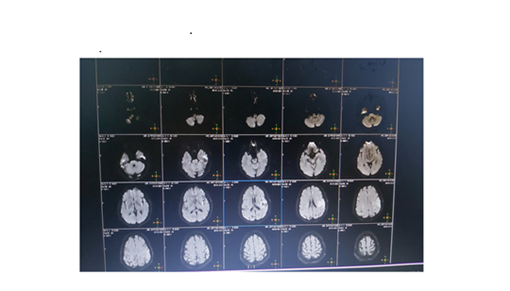
Figure 1. Diffusion MR DAG image
AIS, which ranks first in terms of mortality and morbidity all over the world, is a serious but treatable and preventable disease. However, it is observed that it causes disability in patients who cannot receive appropriate treatment at the appropriate time.
The most common type of stroke in the world is ischaemic stroke. In a study conducted in our country, it was found that 77% of the cases were ischaemic stroke type[3].In recent studies, risk factors for ischaemic stroke were found to be HT, DM, AF, coronary artery disease[4]. AF and HT were also found to be risk factors in our patient.
It is seen that brain computed tomography (CT) is the first neuroimaging modality that can be used to exclude cerebrovascular events in emergency departments after symptoms and physical examination. Non-contrast brain CT scanning excludes haemorrhagic stroke and is an easily accessible method guiding the rapid initiation of treatment after the diagnosis of AI, but has a lower sensitivity [5-6]. On the other hand, magnetic resonance imaging (MRI) has a higher sensitivity to ischaemia compared to CT and is more guiding in predicting long-term prognosis[7].In our patient, brain CT and MRI were performed simultaneously because there was no difference in time and the diagnosis of AIS was made.
In the USA, a multicentre study was conducted by the National Institute of Neurological Disorders and Stroke (NINDS) stroke study group in 1995 and it was shown that IV rt-PA treatment was a safe and effective treatment in patients presenting with acute ischaemic stroke within the first 3 hours and it was included in the guidelines immediately afterwards [8]. In our country, the use of rt-PA (alteplase) was initiated in 2006, and it was recommended to use 0.9 mg/kg, maximum dose 90 mg, for 60 minutes, initially 10% of this dose as a bolus for 1 minute, and after 2018, stroke centres and units were put into operation in many centres [9-10]. Awareness studies have been carried out to take patients to the appropriate centre after the ‘Face, Arm, Speech, Time’ (FAST) test at the scene in order to provide effective treatment to patients with the perception of ‘time is neuron’ with the initiation of treatment with rt-PA (alteplase) in AIS [11]- In another study conducted later, it was observed that rt-PA (alteplase) treatment applied in the first 3-4.5 hours of AIS was effective. However, in the 2010 AHA stroke guideline, it was recommended that iv rt-PA treatment be administered to patients who came within the first 4.5 hours [12].It was observed that there was a relationship between the time of initiation of treatment in AIS and treatment success.The earlier the rt-PA was started, the better the prognosis, especially in those who started within 90 minutes, it was observed that it was more successful and complete recovery was observed in one of every 4.5 patients, and the rate of complete recovery decreased as the time increased [13]. In our case, in accordance with the literature, treatment was started in the first 3 hours and the patient recovered completely and returned to daily life within one month.
AIS is a preventable disease and patients with risk factors should be identified, appropriate treatments should be initiated and followed up. The earlier thrombolytic rt-PA (alteplase) treatment is started after the diagnosis of AI in emergency services, the higher the rate of complete recovery. It should be kept in mind that it will be useful for the teams to take the patients who are thought to have AIS in the foreground to stroke centres with rapid recognition tests in patients taken from the scene of the incident, and necessary studies should be carried out to increase awareness.
Dear Editorial Team, Clinical Medical Reviews and Reports. My experience with the journal was highly positive. The peer-review process was rigorous, constructive, and completed in a timely manner. The reviewers provided valuable comments that helped improve the quality and clarity of our manuscript. The editorial office was professional, responsive, and supportive throughout all stages of the publication process. Communication was clear and efficient, and any questions were addressed promptly. Overall, I found the journal to maintain high scientific standards and an excellent publication workflow. I would be pleased to consider submitting future work to this journal. Best wishes from, Elena Popa.

It was my pleasure to submit my testimonial concerning the Reviewer Board of our Scientific Journal “Brain and Neurological Disorders”. The Reviewers focused on some modifications and their contribution was helpful. The ladies of our Editorial Office were also supported my efforts. It was my honor to have such a co-operation and I am looking forward for more collaboration.

Dear Grace Pierce, Editorial Coordinator of Journal of Clinical Research and Reports, Thank you for the speedy and efficient peer review process. I appreciate the fact that your peer reviewers do not take months to respond like with some other journals. I would also like to thank the editorial office for responding quickly to my questions. It is an excellent journal. I plan to submit more manuscripts in the future. Best wishes from, Robert W. McGee

Dear Grace Pierce, Editorial Coordinator of Journal of Clinical Research and Reports, Working with you and your team on our recent publication in JCRR has been a truly wonderful and enjoyable experience. The responses were prompt, and the reviewers were patient, constructive, and highly professional. One reviewer in particular gave me the feeling that a professor was carefully reading and commenting on my coursework, which was deeply touching. The entire process was straightforward and hassle‑free, with no tedious online forms to complete. I highly recommend this journal. Best wishes from, DR Aibing Rao, Head of R&D

I Appreciate the Opportunity to Share my Experience with the Journal of Clinical Research and Reports. The peer review process was timely and constructive, and the feedback provided helped improve the quality of our manuscript. The editorial office was professional, responsive, and supportive throughout the process, ensuring smooth communication and efficient handling of the submission. Overall, it was a positive experience collaborating with your team.

Dear Mercy Grace, Editorial Coordinator of Obstetrics Gynecology and Reproductive Sciences, We would like to express our gratitude for your help at all stages of publishing and editing the article. The editors of the magazine answer all the necessary questions and help at every stage. We will definitely continue to cooperate and publish other works in the Obstetrics Gynecology and Reproductive Sciences! Best wishes from, Alla Konstantinovna Politova,
