
Case Report | DOI: https://doi.org/10.31579/2768-2757/102
University of Mississippi Medical Center, Division of Plastic Surgery.
*Corresponding Author: John M Sullivan III, University of Mississippi Medical Center, Division of Plastic Surgery.
Citation: John M. Sullivan III, Kacy Benedict, Eric Waetjen, Marc E. Walker, (2024), Acute Free Fibula Reconstruction of a Ballistic Ulna Fracture, Journal of Clinical Surgery and Research, 5(1); DOI:10.31579/2768-2757/102
Copyright: © 2024, John M Sullivan III. This is an open access article distributed under the Creative Commons Attribution License, which permits unrestricted use, distribution, and reproduction in any medium, provided the original work is properly cited.
Received: 14 December 2023 | Accepted: 29 December 2023 | Published: 10 January 2024
Keywords: arm contouring; elbowplasty; brachioplasty; arm lift
Long bone reconstruction is a complicated topic, especially in cases with a large bone deficit where traditional fixation methods would lead to a high rate of non-union requiring further surgical intervention. Our case is of a male with a gunshot wound to his ulna resulting in a 10cm bone defect and a 15x8cm soft tissue defect. Our goal was to maintain a both bone forearm by reconstructing his ulna to maintain forearm pronation and supination. A chimeric free fibula flap was transferred in the acute setting. Vascularized bone transfer in upper extremity reconstruction is typically considered as an operation after fracture non-union. We propose the use of a vascularized bone flap as a first line therapy in certain instances such as the case above. The use of the free fibula flap can provide vascularized bone and softs tissus and allow for optimal recovery while decreasing the need for further operations.
Long bone reconstruction is a complex topic that often climbs many rungs of the reconstruction ladder. Bone reconstruction can be managed with various techniques, all varying in complexity and based on the length of bony reconstruction required. For smaller bone deficits of less than 6 cm, the traditional masquelet technique using a temporary cement spacer followed by staged bone grafting can generally be used provided that the soft tissue coverage is adequate1. Where this technique can be insufficient however is in cases of bone gaps greater than 6 cm, and in cases where there is a poor soft tissue envelope. Additionally, poor candidates for this type of reconstruction include patients with planned postoperative radiation after oncological reconstruction. In these cases, vascularized bone transfer has been shown to be superior to that of the traditional masquelet techinque2. The initial choice for vascularized bone transfer in large bone gaps is a free fibula flap. Advantages of using the fibula over other sources of vascularized bone is not only the significant length of bone that can be harvested, but also the bicortical strength of the fibula. The vascularized fibula flap offers multiple components important for integration and bone healing including osteogenesis, osteoinduction and osteoconduction3. For long bone reconstruction, particularly in the forearm, the fibula is an excellent choice given its similar diameter to the radius and ulna. Prior studies have evaluated use of the free fibula flap as a choice for large bone defects in trauma, and as a choice for secondary reconstruction after failed union of a fracture4-5. However, little has been written at the time of this publication on the use of a free chimeric fibula flap including a soft tissue skin paddle in the acute post traumatic setting for reconstruction of a large long bone deficit. In this case report, we present an acute traumatic wound with a 10 cm ulnar bone gap and soft tissue injury that required a free fibula flap with a large skin paddle for forearm reconstruction of both the osseous and soft tissue components.
The patient, a 42-year-old right hand dominant male, presented to the emergency department following multiple gunshot wounds most notably to his left femur and left forearm.
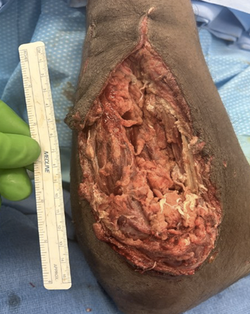
Figure 1: Wound defect prior to reconstruction after multiple debridements illustrating a large soft tissue defect with a long segment of missing ulna.
He was admitted and stabilized initially but taken to the operating room by orthopedic surgery for stabilization of his left femur and irrigation and debridement of his left forearm.
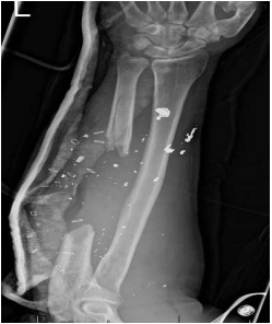
Figure 2: An X-ray of the traumatic limb illustrating a large segment of missing ulna with stable wrist and elbow joints.
Plastic Surgery was consulted intraoperatively for assistance with reconstruction of the resulting 15 x 8 cm soft tissue defect overlying a 10 cm bone deficit in the midshaft of the ulna. A computed tomography scan confirmed that the patient’s major vascular anatomy was intact distal to the injury. A thorough hand exam noted no major nerve or tendinous injuries beyond the fracture. After discussion with the orthopedic surgery team, it was clear that appropriate reconstruction would necessitate both bone and soft tissue replacement in the form of a free flap, given the large defect. The decision was made to proceed with a right free fibula flap, given the additional injury to the patient’s left lower extremity. Given the need for soft tissue coverage over the planned bone flap and fixation, the plan was to proceed with a chimeric free fibula flap including a large skin paddle in the acute setting. An osteocutaneous free fibula was performed with appropriate fixation less than one week following the initial trauma.
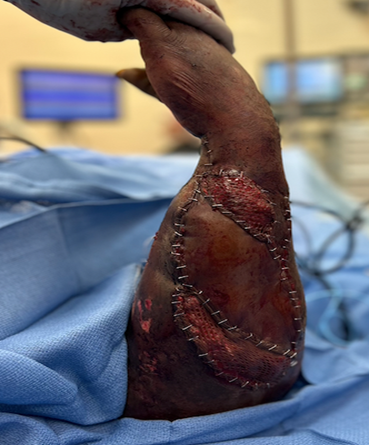
Figure 3: Intra-operative photo of free fibula flap after placing it in the ulna defect and using the skin paddle to assist with soft tissue coverage over the flap. The remaining soft tissue was skin grafted.
After appropriate debridement of the forearm wound, vertical osteotomies of the remaining native ulna were performed to create a landing zone for the free flap. The fibula was then affixed to the proximal and distal ends of the ulna with low profile reconstruction plates using monocortical bone screws on the fibula flap and bicortical bone screws to the native proximal and distal ulna. All the implanted hardware was appropriately covered with the fibula flap skin paddle and small skin grafts were used to cover the remaining soft tissue wound of the upper extremity and for closure of the fibula flap donor site. Radiographs following dressing removal at one week postoperatively showed good approximation of the free fibula flap interposed in the defect of the ulna.
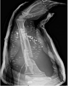
Figure 4: A postoperative X-ray that illustrates adequate inset of the free fibula flap into the ulna defect and plated appropriately with good boney contact proximally and distally.
Forearm reconstruction is a challenging topic and one that requires foresight into the end goals of the patient. For our patient, supination and pronation of the forearm was an important reconstructive goal for him to achieve. With this in mind, and given his young age, we wanted to provide him with the best opportunity for motion by reconstructing his ulna rather than performing wrist arthrodesis and one-bone forearm reconstruction6. One option for reconstruction to maintain a both-bone forearm would have been to attempt a masquelet procedure using a large dorsal plate and bone cement followed by serial bone grafting. However, given his poor soft tissue envelope, we were concerned that the hardware would quickly become exposed and result in failure of this technique. From this, we considered use of a fasciocutaneous free flap combined with the masquelet as surgically this could be staged. However, this would require a return to the operating room and possibly still necessitate vascularized bone given the large size of the osseous gap.
In response to these issues, we decided that if a free flap was going to be required for reconstruction, then the best course of action would be to perform one in which we could bring bone as well as soft tissue. While there are a few options for vascularized bone reconstruction, only one allows for a strong bicortical fixation with a similar diameter for the ulna. We elected to perform an acute free vascularized fibula flap with individual proximal and distal fixation. This allowed us to bring not only a large skin paddle but also a long segment of bone. The fibula was selected over other osteocutaneous flaps as it would provide a bony segment with similar diameter and strength as the native ulna. This plan addressed many of the patient’s main concerns by offering him the ability to maintain pronation and supination of the forearm and allowing him to have a “one and done” procedure which ultimately decreased his hospital time. Additionally, it provided not only vascularized bone, but the soft tissue required to adequately cover the bony reconstruction. Based on our experience, consideration should be placed on performing an acute free fibula in select patient where bone as well as soft tissue is required for reconstruction with the additional benefit of providing a quicker surgical recovery.
The vascularized free fibula flap is an excellent option for complex long bone reconstruction and has been well described as a suitable size match for the radius and ulna.3 As illustrated in our case, the use of this as a first line therapy in the acute setting rather than in a delayed fashion after a nonunion or an attempted masquelet should be considered. Especially if soft tissue reconstruction is required as the flap can be harvested in a chimeric fashion to include both a skin paddle and a vascularized bone segment. This is a useful reconstructive technique to decrease the total number of surgeries required which aids in reducing the total duration of inpatient stay. Written informed consent was obtained from the patient for publication of this case report and accompanying identifying images.
Dear Editorial Team, Clinical Medical Reviews and Reports. My experience with the journal was highly positive. The peer-review process was rigorous, constructive, and completed in a timely manner. The reviewers provided valuable comments that helped improve the quality and clarity of our manuscript. The editorial office was professional, responsive, and supportive throughout all stages of the publication process. Communication was clear and efficient, and any questions were addressed promptly. Overall, I found the journal to maintain high scientific standards and an excellent publication workflow. I would be pleased to consider submitting future work to this journal. Best wishes from, Elena Popa.

It was my pleasure to submit my testimonial concerning the Reviewer Board of our Scientific Journal “Brain and Neurological Disorders”. The Reviewers focused on some modifications and their contribution was helpful. The ladies of our Editorial Office were also supported my efforts. It was my honor to have such a co-operation and I am looking forward for more collaboration.

Dear Grace Pierce, Editorial Coordinator of Journal of Clinical Research and Reports, Thank you for the speedy and efficient peer review process. I appreciate the fact that your peer reviewers do not take months to respond like with some other journals. I would also like to thank the editorial office for responding quickly to my questions. It is an excellent journal. I plan to submit more manuscripts in the future. Best wishes from, Robert W. McGee

Dear Grace Pierce, Editorial Coordinator of Journal of Clinical Research and Reports, Working with you and your team on our recent publication in JCRR has been a truly wonderful and enjoyable experience. The responses were prompt, and the reviewers were patient, constructive, and highly professional. One reviewer in particular gave me the feeling that a professor was carefully reading and commenting on my coursework, which was deeply touching. The entire process was straightforward and hassle‑free, with no tedious online forms to complete. I highly recommend this journal. Best wishes from, DR Aibing Rao, Head of R&D

I Appreciate the Opportunity to Share my Experience with the Journal of Clinical Research and Reports. The peer review process was timely and constructive, and the feedback provided helped improve the quality of our manuscript. The editorial office was professional, responsive, and supportive throughout the process, ensuring smooth communication and efficient handling of the submission. Overall, it was a positive experience collaborating with your team.

Dear Mercy Grace, Editorial Coordinator of Obstetrics Gynecology and Reproductive Sciences, We would like to express our gratitude for your help at all stages of publishing and editing the article. The editors of the magazine answer all the necessary questions and help at every stage. We will definitely continue to cooperate and publish other works in the Obstetrics Gynecology and Reproductive Sciences! Best wishes from, Alla Konstantinovna Politova,
