
Review Article | DOI: https://doi.org/10.31579/2643-6612/016
1Department of Orthodontics, Terna Dental College,Nerul Navi Mumbai
2 Department of Orthodontics, Yogita Dental College, Khed, Ratnagiri, Mumbai
*Corresponding Author: Rohit Kulshrestha, Department of Orthodontics, Terna Dental College, and Nerul Navi Mumbai
Citation: Rohit K, Snehal B, Pavankumar V, Chetan P, Vinay U, Balagangadhar(2019) Accelerated Orthodontics: A Review. J Dentistry And Oral Maxillofacial Surgery 1(1); Doi: 10.31579/2643-6612/016
Copyright: ©2019 Rohit Kulshrestha, This is an open-access article distributed under the terms of The Creative Commons. Attribution License, which permits unrestricted use, distribution, and reproduction in any medium, provided the original author and source are credited.
Received: 05 December 2019 | Accepted: 19 December 2019 | Published: 30 December 2019
Keywords: accelerated orthodontic tooth movement, corticotomy, peizocision, micro-osteoperforation(MOPs).
Nowadays, there is an increased tendency for researches to focus on accelerating methods for tooth movement due to the greater demand for adults for a shorter orthodontic treatment duration. Unfortunately, long orthodontic treatment time has many disadvantages like higher predisposition to caries, gingival recession, and root resorption. This increases the demand to increase tooth movement with the least possible disadvantages. Several modalities have been reported for accelerating the tooth movement. Thus, accelerating orthodontic tooth movement and the resulting shortening of the treatment time would be quite beneficial.
There is an increased demand for fast orthodontic treatment, especially by adult patients. This has led researchers to focus on accelerated tooth movement with the aim of reducing treatment duration while maintaining treatment efficiency. The approach that researchers select to accelerate the rate of movement depends on interpretation of the data on the biology of tooth movement.
Orthodontic tooth movement occurs because of a mechanical stimuli which is sequenced by remodelling of the alveolar bone and periodontal ligament (PDL). Bone remodelling is a process of both bone resorption on the pressure site and bone formation on the tension site [1]. Orthodontic tooth movement can be controlled by the amount of the applied force and the biological responses from the PDL [2]. The force applied on the teeth will cause changes in the microenvironment around the PDL due to alterations of blood flow, leading to the secretion.
Three types of bone cells play a significant role in the biology of tooth movement: osteoblasts, osteocytes, and osteoclasts. Osteoblasts are mononuclear cells found along bone surfaces. They are derived from mesenchymal stem cells in the bone marrow and synthesize collagenous and non-collagenous proteins that comprise the organic bone matrix, known as osteoid. Inactive osteoblasts, particularly in the adult skeleton, are called bone-lining cells. These cells are quiescent until growth factors or other anabolic stimuli induce their proliferation and differentiation into cuboidal osteoblasts. Osteoblasts are the main cells participating in the anabolic phase of orthodontic tooth movement with a limited role during the catabolic phase.
The mechanism by which mechanical stimulation activates osteocytes is not clear. Bone loading under physiologic condition results in strain, or deformation, in the bone matrix and the osteocyte lacunae and canaliculi. Some investigators suggest that it is the strain magnitude in the matrix, rather than in the lacunae or canaliculi, that triggers bone remodeling. These responses to mechanical load activate osteocytes to secrete key factors, such as prostaglandins, nitric oxide, or insulin-like growth factors (IGFs), which then activate osteoclasts and osteoblasts in a tightly synchronized biological phenomenon called bone remodeling.
Osteoclasts control the rate of bone resorption during orthodontic treatment and, therefore, the rate of tooth movement. However, osteoclasts do not function independently. In fact, they require signals from several other cell types for their maturation, activation, and ability to perform targeted, site-specific bone resorption. The consequences of unregulated osteoclast activation would be catastrophic as bone resorption would proceed unchecked producing weakened bone and fractures. Consequently, osteoclasts cannot be direct targets of orthodontic forces. Instead, the upstream events that control osteoclast formation and activation must be the main targets.
Methods of Acceleration:
The basis for clinical procedures such as corticotomy-assisted orthodontics, piezocision-aided orthodontics, and surgery-first orthodontics is the direct injury to the both alveolar bones (maxillary arch and mandibular arch) AOTM by inducing regional acceleratory phenomenon (RAP), as a wound-healing process [3,4]. A surgical technique was introduced by Bichalmyr [5] for faster correction of severe maxillary protrusion with available orthodontic appliances. Wedges of alveolar bone were removed to decrease the volume of the bone through the radicular parts of the maxillary teeth in anterior region. Kole [6] suggested a technique of creating bony blocks (bone-teeth unit) through the corticotomy to enhance the faster tooth movement. For the next fifty years this concept prevailed until Wilcko and co-workers [7], reported a transient demineralization -remineralization procedure enchanting after corticotomy, which was termed as a periodontally accelerated osteogenic orthodontics (PAOO). The PAOO is an amalgamation of selective decortications and facilitated orthodontic techniques along with alveolar augmentation. This technique shortens the treatment time to 33% the time of conventional treatment duration in orthodontics. This concept was based on a similar technique, as regional acceleratory phenomena (RAP). This method is a local response to a lethal stimulus which describes a process of tissue formation faster than the usual local regeneration process. Enhancing a variety of healing stages, this RAP makes healing occur 2-10 times earlier than regular healing [8]. However, this is an old technique and very invasive hence, this was not accepted by all patients. Hence, latest procedures like corticision, piezosurgery, fiberotomy and microosteoperforations had demonstrated.
Bone is surgically wounded so as to initiate a localized inflammatory response. The presence of cytokines and chemokines through prostaglandin E2 pathway and the RANK/ RANKL pathway causes differentiation of osteoclasts which leads to bone resorption and thus AOTM is possible. It has been reported that this effect lasts for 4 months and this method needs to be repeated, in case faster tooth movement further if required.
Inter-septal alveolar surgery: Inter-septal alveolar surgery or distraction osteogenesis involves controlled and gradual displacement of surgically created fractures which is termed as sub-periosteal osteotomy by incremental traction that results in simultaneous expansion of soft tissue and the surrounding alveolar bone volume due to mechanical stretching of the osteotomy site. It is divided into the distraction of the dentoalveolar bone or distraction of periodontal ligament [9]. (fig. 1).
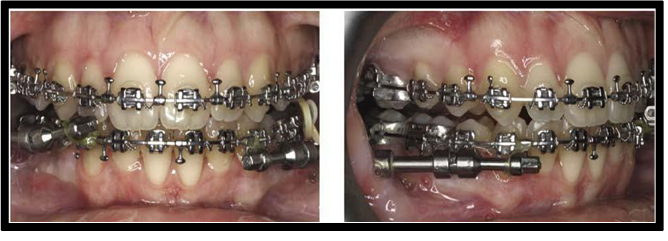
Procedure: At the time extraction of first premolars the inter-septal bone distal to the canine is undermined surgically. Eventually resistance on the pressure site will be decreased. Bone distal to canine undermined inter-septally by 1 to 1.5mm. Based on inter-septal alveolar surgery, the compact bone is replaced by the woven bone, and tooth movement is easier and quicker due to reduced resistance of the bone. These rapid movements are found to be achieved during the initial phases of tooth movement, especially in the first week. Rapid canine distraction of the dento-alveolar bone may be performed by the same principle of the distraction of periodontal ligament with the addition of more dissection and osteotomies performed at the vestibule, in some cases. Clinical trials on humans showed that this technique would reduce the resistance in the pathway of canine movement more effectively during orthodontic treatment. Among the studies reported on inter-septal alveolar surgery two were cross sectional studies and rest were randomized clinical trials (RCTs).
Corticotomy: A corticotomy is defined as a surgical procedure whereby only the cortical bone is cut, perforated, or mechanically altered without any alteration in the medullary bone. This is performed without the involvement of medullary bone unlike osteotomies which involve the entire thickness of bone [10].
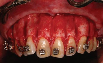
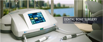
Procedure: Elevation of full thickness mucoperiosteal flaps of both buccal and/or lingual region. Positioning the corticotomy cuts using piezosurgical armamentarium or micromotor under irrigation and it is followed by placement of a graft material, in required sites to enhance the thickness of the bone [10].
Advantages: Bone can be augmented and periodontal defects would be avoided. Minimal changes in the periodontal attachment apparatus. Shorter treatment time. Less root resorption. Various authors performed clinical trials using corticotomy assisted canine retraction and found reduction in treatment time by 28%-33% and a 2-3-fold increase in velocity of tooth movement when compared with conventional OTM on control side.
Disadvantages: Expensive and comparatively invasive procedure. May cause post-operative pain and swelling.
Corticision: Kim and co-workers established a technique with minimal surgical intervention called corticision which is also called as minimally invasive rapid orthodontics (MIRO). Corticision was initiated as a supplemental dento-alveolar surgery in orthodontic therapy to achieve AOTM with minimal surgical intervention[11].
Procedure: Separation of the inter-proximal cortices with a reinforced scalpel is used as a thin chisel and a mallet transmucosally without reflecting a flap. With 45°-60° an inclination to the gingiva at the long axis of the canine a reinforced surgical blade with a minimum thickness of 400 μm should be located on the inter-radicular attachment. In order to preserve the alveolar crest, the surgical injury should be 2 mm from the papillary gingival margin and 1 mm beyond the mucogingival junction. The blade should be pulled out by a swing motion. Clinical studies were conducted on humans and animals and concluded corticision effectively accelerates the tooth movement similar to corticotomy and is more advantageous because of its less invasiveness. Among the published studies on corticision two were case control studies and other was case series.
Piezocision: This is a minimally invasive procedure involves flapless in combining piezosurgical cortical micro-incisions with selective tunneling that allows for soft-tissue or bone grafting. Vercelotti and Podesta established the use of piezosurgery instead of burs, in conjunction with the conventional flap elevations to create an environment conducive for the rapid tooth movement. This technique is quite invasive as it requires extensive flap elevation and osseous surgeries, with post-surgical discomfort. This technique has not been widely accepted by patient community. Subsequently, Dibart introduced piezocision with less invasiveness to this procedure.[12]
Procedure: This is a combination of microincisions limited to the buccal gingiva that allows the use of a piezoelectric knife to give osseous cuts to the buccal cortex and initiate the RAP without involving palatal or lingual cortex. The procedure allows for rapid tooth movement without the downside of an extensive and traumatic surgical approach while maintaining the clinical benefit of a soft-tissue or grafting concomitant with a tunnel approach. Dibart and co-workers[12] established a minimally invasive flapless procedure, combining micro incisions, piezoelectric incisions and selective tunneling that allows for hard- or soft-tissue grafting. They concluded that piezocision allows a rapid correction without the drawbacks of traumatic conventional corticotomy procedures in severe malocclusion cases. They later combined this technique with invisalign and found to be more effective and esthetic.
Microosteoperforations (MOPs): A device used for this method is called as PropelTM, which was launched by Propel Orthodontics. It reduces the invasive nature of surgical irritation of bone. This procedure was initially popularized as alveocentesis, which literally means puncturing of bone. The device has an adjustable depth dial at 0mm, 3mm, 5mm, and 7mm of tip depth and an indicating arrow on the driver body. This device comes as ready-to-use sterile disposable device.
Procedure: A soft tissue flap was raised in the premolar and molar region and small perforations of about 0.25 mm are made using a round bur and hand piece through the cortical bone. 1-3 micro-osteoperforations are to be done depending on proximity of anatomical structures. Perforations can be made on buccal or lingual side of both maxillary and mandibular arch in linear or triangular patterns. Two randomized control trial studies were reported on microosteoperforations among these one was animal study and other was a human trial [13].
Contemporary status of surgical methods: Surgical methods are invasive procedures and hence, patient cooperation is much needed. Inter-septal alveolar surgery, corticotomy and corticision are more invasive and expensive with needed surgical cuts and osteotomies. Post-operative complications are sometimes present with pain, swelling and patient discomfort. Recent techniques such as piezocision and microosteoperforations are less invasive with comparatively less complications than the previously used procedures. But more research is required to be done in using those techniques for accelerating the orthodontic tooth movement.
Device assisted therapy or mechanical stimulation methods:
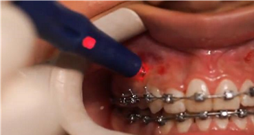
There are various methods used to accelerate tooth movement like direct electric currents, resonance vibration, low level laser therapy, static magnetic field, and pulsed electromagnetic field. The concept of applying orthodontic forces causes bone bending (bone bending theory) and bioelectrical potential gave an idea of using physical approaches.The bioelectrical potential is created when there is application of discontinuous forces, which leads to the idea of trying cyclic forces and vibrations.
Direct electric current: Electrical current has been tested experimentally on the animal models and have shown good results with accelerating orthodontic tooth movement. Direct current or electrical currents generated piezoelectrically thereby enhance the orthodontic tooth movement.
Procedure: An electric appliance that provides direct electric current was placed in the extracted tooth region, generated bio electric potentials causing local responses and acceleration of bone modelling. This procedure was performed by some researchers on living animals and found to be effective in tooth movement. Subsequently, Kim performed a clinical trial on humans and found 30
Acceleration of tooth movement while orthodontic treatment is of increasing demand now a days because of patient’s interest to get the treatment completed in less span of time and to decrease the number of visits. And even the adult orthodontics has gained more demands as the adult patients are increasing day by day for the orthodontic treatment. Surgical techniques are more invasive and costly but are more beneficial with fewer side effects. Hence, recent techniques such as piezocision, micro-osteoperforations has the more demand in future. Less invasive surgical techniques can be safely used to accelerate tooth movement with increased patient compliance. Device assisted therapy is also of high demand but there is a need for further studies about the proper device being used and how far it is useful. Pharmacological methods have more side effects and hence most of them are still in experimental stage. Only limited human trails are available. Accelerating orthodontic techniques can be highly useful for fastening the treatment as in every technique being used; there is increased rate of tooth movement and hence decreasing the treatment time.
Dear Editorial Team, Clinical Medical Reviews and Reports. My experience with the journal was highly positive. The peer-review process was rigorous, constructive, and completed in a timely manner. The reviewers provided valuable comments that helped improve the quality and clarity of our manuscript. The editorial office was professional, responsive, and supportive throughout all stages of the publication process. Communication was clear and efficient, and any questions were addressed promptly. Overall, I found the journal to maintain high scientific standards and an excellent publication workflow. I would be pleased to consider submitting future work to this journal. Best wishes from, Elena Popa.

It was my pleasure to submit my testimonial concerning the Reviewer Board of our Scientific Journal “Brain and Neurological Disorders”. The Reviewers focused on some modifications and their contribution was helpful. The ladies of our Editorial Office were also supported my efforts. It was my honor to have such a co-operation and I am looking forward for more collaboration.

Dear Grace Pierce, Editorial Coordinator of Journal of Clinical Research and Reports, Thank you for the speedy and efficient peer review process. I appreciate the fact that your peer reviewers do not take months to respond like with some other journals. I would also like to thank the editorial office for responding quickly to my questions. It is an excellent journal. I plan to submit more manuscripts in the future. Best wishes from, Robert W. McGee

Dear Grace Pierce, Editorial Coordinator of Journal of Clinical Research and Reports, Working with you and your team on our recent publication in JCRR has been a truly wonderful and enjoyable experience. The responses were prompt, and the reviewers were patient, constructive, and highly professional. One reviewer in particular gave me the feeling that a professor was carefully reading and commenting on my coursework, which was deeply touching. The entire process was straightforward and hassle‑free, with no tedious online forms to complete. I highly recommend this journal. Best wishes from, DR Aibing Rao, Head of R&D

I Appreciate the Opportunity to Share my Experience with the Journal of Clinical Research and Reports. The peer review process was timely and constructive, and the feedback provided helped improve the quality of our manuscript. The editorial office was professional, responsive, and supportive throughout the process, ensuring smooth communication and efficient handling of the submission. Overall, it was a positive experience collaborating with your team.

Dear Mercy Grace, Editorial Coordinator of Obstetrics Gynecology and Reproductive Sciences, We would like to express our gratitude for your help at all stages of publishing and editing the article. The editors of the magazine answer all the necessary questions and help at every stage. We will definitely continue to cooperate and publish other works in the Obstetrics Gynecology and Reproductive Sciences! Best wishes from, Alla Konstantinovna Politova,
