

Research Article | DOI: https://doi.org/10.31579/2641-5194/029
Department of Radiology, Erzincan Binali Yildirim University, School of Medicine, Erzincan, Turkey.
*Corresponding Author: Ozlem Kadirhan, Department of Radiology, Erzincan Binali Yildirim University, School of Medicine, Erzincan, Turkey.
Citation: Kadirhan O. Aydin S. , Kantarci M. (2021) A Rare Cause of Acute Abdomen in Children: Spontaneous Rupture of Choledochal Cyst Causing Peritonitis. J.Gastroenterology Pancreatology and Hepatobilary Disorders 5(3) DOI:10.31579/2641-5194/029
Copyright: © 2021, Ozlem Kadirhan,This is an open access article distributed under the Creative Commons Attribution License, which permits unrestricted use, distribution, and reproduction in any medium, provided the original work is properly cited.
Received: 07 May 2021 | Accepted: 11 May 2021 | Published: 17 June 2021
Keywords: choledochal cyst; rupture; bilious ascites; peritonitis; roux-en-Y hepaticojejunostomy
Spontaneous rupture of the common bile duct cyst is a rare cause of acute abdomen in children.
In our case, we present a 3-year-old patient with no history of trauma, whose diagnosis was confirmed by laparotomy, with a possible suspected common bile duct cyst due to massive ascites and a cystic lesion in the portal area.
Although common bile duct cysts are mostly asymptomatic, various studies have shown that malignant transformation can cause important complications such as pancreatitis.Numerous causes have been suggested in the etiopathogenesis of rupture, such as parietal necrosis associated with pancreatic reflux irritation.Since rupture of cysts may require urgent laparotomy such as biliary peritonitis, it is important to know imaging findings that may be warning for early diagnosis.
Because of the increased risk of cholangiocarcinoma after cyst excision, cholecystectomy and Roux-en-Y hepaticojejunostomy are the most common treatment procedures.
1. Conception - OK, SA
2. Design - OK, SA
3. Supervision - SA, MK
4. Fundings - None
5. Materials - OK, SA
6. Data Collection and/or Processing - OK, SA, MK
7. Analysis and/or Interpretation - SA, MK
8. Literature Review - OK, SA
9. Writing - OK, SA
10. Critical Review Contribution Type-SA, MK
Common bile duct cysts are mostly congenital abnormal enlargement of the biliary tract .In America and Europe, the incidence of common bile duct cyst disease ranges from 1 in 100,000 to 1 in 150,000 live births, and from 1 in 5,000 to 1 in 1,000 live births in Asia [1, 2] . Symptoms most commonly associated with common bile duct cysts include right upper quadrant pain, jaundice,vomiting and sometimes palpable mass ,and the rate of these symptoms ranges from 22 to 47% in one recent study [2, 3] .Common bile duct cysts have potentially life-threatening complications.Some patients may present with rupture of the cyst itself, which is a rare cause of acute abdomen, and secondary biliary peritonitis [4]. In a large case series of 1433, the number of patients with bile peritonitis due to rupture of the common bile duct cyst was 26 (1.81%) [5]. Since this situation requires urgent laparotomy, early diagnosis is important.
We present a case report of spontaneous choledochal cyst rupture at the age of 3 years without a history of trauma complicated by peritonitis.
A 3-year-old male patient was brought to our hospital with complaints of fever, vomiting, diarrhea, abdominal pain and abdominal distension that had been increasing for four days. There was no history of preceeding trauma. On physical examination,diffuse abdominal tenderness, decreased bowel sounds ,fever (38,5oC) , and jaundice was present. Blood tests showed leukocytosis (22,300 cells/mm3), increased C-reactive protein( 127 mg/ml) ; total bilirubin (15.2 mg/dL) , alkaline phosphatase (400 IU/L)and amylase(380 U/l ) levels.
Imaging Findings were the following: Abdominal ultrasound(USG) was performed.Little information could be acquired because of abdominal gas and tenderness;massive ascites was defined in abdomen and pelvis. Computed tomography (CT) examination then performed; a cystic structure with a diameter of 3 cm was detected beside of the pancreatic head and choledoc.Also,dilated bowel loops, and a large amount of turbid free fluid is seen. In addition, there was an increase in size compared to normal in the pancreas.
In the paracentesis we performed later, we found that there was a high level of bilirubin in the intraabdominal fluid.The patient underwent an emergency abdominal surgery with laparotomy. On laparoscopy , a 3-cm cystic mass was found infero laterally to the gallbladder and it was partially embedded in the subhepatic fossa.There was a generalized biliary peritonitis related to the perforation of the cystic lesion . The lesion was associated with the common bile duct via stalk. Intraoperative cholangiography showed an isolated diverticulum protruding from the common bile duct.
It was learned that the patient was admitted to the pediatric surgery service, and that broad spectrum antibiotherapy was initiated first, and a T-tube was placed in order to provide biliary drainage with emergency surgery. Roux-en-Y hepaticojejunostomy was performed together with total cystectomy at the 10th week in the patient whose general condition improved on the 9th day after emergency surgery.
It was learned that he was discharged on the tenth day after the definitive surgery without complications. In the intermittent 15-month follow-ups, it was reported that the general condition of the patient was good and no complications developed yet.
Figures
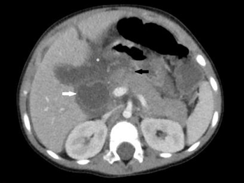
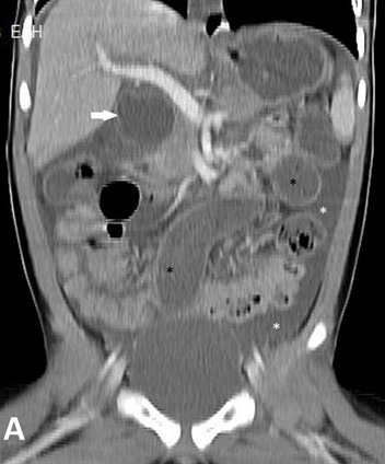

A choledochal cyst (CC) is a rare congenital cystic dilation of the extrahepatic and intrahepatic biliary tract, most common in females of Asian descent [6]. It has been shown in different studies that the incidence is higher in female [3, 4, 7] .
In the series of 74 cases compiled by Hsu et al, it was revealed that the most common forms of presentation were abdominal pain, vomiting, anorexia, and jaundice [3].Although these cysts are mostly asymptomatic at first, it has been shown as a result of various studies that they can cause complications such as malignant transformation, cholangitis, pancreatitis and stone formation [4, 8] .Spontaneous rupture of the cyst is very critical and causes biliary peritonitis that requires urgent laparotomy [4].
The etiopathogenesis of perforation may be related to a variety of mechanisms, such as parietal necrosis due to thrombosis of intramural vessels or associated with pancreatic reflux irritation, parietal infection favored by ductal stasis, and ductal highpressure over a barrier. It is still not possible to give a precise reason for this [9] .
The classification of choledochal cysts has been divided into six types by Todani et al [10].In our case, an isolated diverticulum protruding from the wall of the common bile duct was seen and thediagnosis was compatible with type 2 common bile duct cyst.
In a patient who does not have a prior diagnosis or suspicion of common bile duct cyst rupture, the correct diagnosis of this condition is difficult even with laparotomy because the cyst usually shrinks after perforation due to collapse and the usual location of the perforation is highly in the posterior wall.Ultrasonography (USG), computed tomography (CT), intravenous cholangio graphy (IVC) and percutaneous transhepatic cholangiography (PTK), ERCP, magnetic resonance cholangiography (MRCP) may contribute to the diagnosis of choledochal cyst [5, 11, 12]. Ultrasound is the first imaging tool be cause of its noninvasivity and low cost, which is there as on for its frequent use, but other imaging methods are used for various reasons to reach the diagnosis
Using imaging methods such as ultrasound and computed tomography in a patient with acute abdominal symptoms Intraabdominal free fluid, non-dilated bile ducts or gall bladder may be supportive in the preoperative diagnosis of biliary peritonitis by showing a pseudocyst or decompressed cystic lesion in the portal area [13, 14] . Although paracentesis is the fastest way to characterize ascites, contamination risk should be considered [13] .
There is still no single definitive procedure for the treatment of choledochal cyst rupture, but in the presence of a perforated common bile duct cyst, a single-stage biliary cholecystectomy is usually performed with a Roux-en-Y hepaticojejunostomy to minimize the possibility of developing cholangiocarcinoma later in life [15] .
Spontaneous common bile duct cyst rupture is a rare cause of acute abdomen in children and requires urgent surgical treatment. In the patient with acute abdominal findings, some laboratory tests and especially imaging findings may enable us to consider spontaneous rupture of common bile duct cyst in differential diagnosis. Early diagnosis and treatment of spontaneous common bile duct cyst rupture is important in preventing various complications, especially malignant transformation.
Dear Editorial Team, Clinical Medical Reviews and Reports. My experience with the journal was highly positive. The peer-review process was rigorous, constructive, and completed in a timely manner. The reviewers provided valuable comments that helped improve the quality and clarity of our manuscript. The editorial office was professional, responsive, and supportive throughout all stages of the publication process. Communication was clear and efficient, and any questions were addressed promptly. Overall, I found the journal to maintain high scientific standards and an excellent publication workflow. I would be pleased to consider submitting future work to this journal. Best wishes from, Elena Popa.

It was my pleasure to submit my testimonial concerning the Reviewer Board of our Scientific Journal “Brain and Neurological Disorders”. The Reviewers focused on some modifications and their contribution was helpful. The ladies of our Editorial Office were also supported my efforts. It was my honor to have such a co-operation and I am looking forward for more collaboration.

Dear Grace Pierce, Editorial Coordinator of Journal of Clinical Research and Reports, Thank you for the speedy and efficient peer review process. I appreciate the fact that your peer reviewers do not take months to respond like with some other journals. I would also like to thank the editorial office for responding quickly to my questions. It is an excellent journal. I plan to submit more manuscripts in the future. Best wishes from, Robert W. McGee

Dear Grace Pierce, Editorial Coordinator of Journal of Clinical Research and Reports, Working with you and your team on our recent publication in JCRR has been a truly wonderful and enjoyable experience. The responses were prompt, and the reviewers were patient, constructive, and highly professional. One reviewer in particular gave me the feeling that a professor was carefully reading and commenting on my coursework, which was deeply touching. The entire process was straightforward and hassle‑free, with no tedious online forms to complete. I highly recommend this journal. Best wishes from, DR Aibing Rao, Head of R&D

I Appreciate the Opportunity to Share my Experience with the Journal of Clinical Research and Reports. The peer review process was timely and constructive, and the feedback provided helped improve the quality of our manuscript. The editorial office was professional, responsive, and supportive throughout the process, ensuring smooth communication and efficient handling of the submission. Overall, it was a positive experience collaborating with your team.

Dear Mercy Grace, Editorial Coordinator of Obstetrics Gynecology and Reproductive Sciences, We would like to express our gratitude for your help at all stages of publishing and editing the article. The editors of the magazine answer all the necessary questions and help at every stage. We will definitely continue to cooperate and publish other works in the Obstetrics Gynecology and Reproductive Sciences! Best wishes from, Alla Konstantinovna Politova,
