
Case Report | DOI: https://doi.org/10.31579/2578-8965/056
Associate Professor of Medicine, Ad-din Women’s Medical College Hospital, Dhaka, Bangladesh.
*Corresponding Author: Richmond Ronald Gomes ,Associate Professor of Medicine, Ad-din Women’s Medical College Hospital, Dhaka, Bangladesh.
Citation: Richmond R. Gomes (2021) A Rare Case of Giant Ovarian Serous Cystadenoma presenting as Psuedo-Meigs Syndrome. J Obstetrics Gynecology and Reproductive Sciences 5(2): DOI: 10.31579/2578-8965/056
Copyright: © 2021, Richmond Ronald Gomes, This is an open access article distributed under the Creative Commons Attribution License, which permits unrestricted use, distribution, and reproduction in any medium, provided the original work is properly cited.
Received: 12 January 2021 | Accepted: 01 March 2021 | Published: 12 March 2021
Keywords: meigs, pseudo-meigs; serous cystadenoma; hydrothorax; ascites
Meigs’ syndrome is a rare condition characterized by the presence of a benign fibroma of the ovary, ascites and pleural effusion. Other benign cysts of the ovary (such as struma ovarii, mucinous cystadenoma, serous cystadenoma and teratomas), leiomyoma of the uterus, and secondary metastatic tumours to ovary if associated with hydro thorax and ascites are referred to as ‘Pseudo‐Meigs” syndrome. It very uncommon and diagnosis is made difficult by symptoms that usually mimic disseminated malignancy or tuberculosis. The gold standard treatment is laparotomy and, by definition of the syndrome, after tumor removal, the symptoms resolves and the patients become asymptomatic. We presented an 18 years old girl with giant ovarian serous cystadenoma with associated pseudo-meigs syndrome, successfully managed in a low resources setting.
Meigs' syndrome is diagnosed based on a triad of an ovarian fibroma, pleural effusion and ascites. It resolves spontaneously after the resection of the fibroma [1]. In 1852, Blin published the description of an ovarian fibroma with abdominal effusion in the Société de Biology de Paris (cited by Lallemand) [2]. A Demons of Bordeaux, France, gave a report to the Société de Chirurgie de Paris in 1887, that nine of 50 patients with ovarian cysts were cured of their ascites and hydrothorax by removal of the adnexal cyst. In 1937, Joe Vincent Meigs (1892–1963), an American professor of the Harvard Medical School of Gynaecology drew widespread attention of the medical profession to the syndrome [3]. Meigs used seven cases to highlight the association between a fibroma of the ovary, ascites, and hydrothorax. It was coined as Meigs' syndrome in 1937 by Rhodes and Terrell [4].
The following four characteristics were selected by Meigs in 1945 to define the syndrome:
Other benign cysts of the ovary (such as struma ovarii, mucinous cystadenoma, serous cystadenoma and teratomas), leiomyoma of the uterus, and secondary metastatic tumours to ovary if associated with hydro thorax are referred to as ‘Pseudo‐Meigs” syndrome. .[6]
An atypical case of Meigs' syndrome was reported in 1990 by Martin, et al. .[7] presenting as bilateral sanguineous pleural effusion without ascites in a woman with a granulosa cell tumor.
We presented a giant ovarian serous cystadenoma with associated pseudo-Meigs syndrome, successfully managed in a low resources setting.
Ms. Sharmin, a 18 years old unmarried student presented to the department of Medicine, Ad-din Women’s Medical College Hospital with the complaints of gradual distension of abdomen with dull aching lower abdominal pain and feeling of shortness of breath for last 2 months. She denied any fever, chest pain, cough, weight loss or altered bowel habit. Her menstrual history was normal. She had no history of exposure with patient with active tuberculosis. She was vaccinated as per EPI schedule. Past medical and surgical history were insignificant. On general examination she was of average build with mild pallor. Jaundice, cyanosis, clubbing and edema were absent. Her pulse rate was 88/ minute, blood pressure - 110/76 mm of Hg, respiratory rate 24/minute and temperature normal. Breast examination revealed no abnormalities. Respiratory system examination revealed a stony dullness over right chest wall from 6th intercostal space downwards. Breath sounds were diminished on the right side over the same area. There were no crepitations or rhonchi. The cardiovascular system examination showed no adventitious sounds with S1 and S2 being normally audible. Abdominal inspection revealed fullness of whole abdomen without any prominent veins or distortion of the umbilicus. No mass could be palpated but there was tenderness in the right iliac fossa and part of the hypogastrium. Liver, spleen, and inguinal lymph nodes were not palpable. On percussion there was shifting dullness due to free fluid. Auscultation revealed normal intestinal peristaltic sounds. Vaginal examination showed a right sided solid mass of about 20 cm size separate from the normal sized uterus. Other systemic examinations revealed no abnormalities.
Routine investigations including complete hemogram, blood sugar, blood urea, s. creatinine, s. albumin, s. amylase, s .lipase, liver function tests and electrolytes, viral markers (HBsAg, Anti HCV) were within normal limits. Chest x-ray showed right sided pleural effusion (Figure 1). MRI of abdomen revealed huge cystic lesion measuring about 18cm×19cm×7cm, arising from right adnexal region with moderate ascites and moderate right sided pleural effusion (Figure 2, figure 3 and figure 4). Paracentesis was done. Ascitic fluid study revealed exudative fluid with protein 3.9 gm/dl. Gram stain and malignant cells in ascetic fluid were negative. ADA (adenosine deaminase) was normal of 8.68 U/L (normal <25>
Histopathology revealed cyst wall lined by cuboidal epithelium supported by fibrocollagenous tissue suggestive of serous cystadenoma with no evidence of malignancy (Figure-6). Attached fallopian tube was unremarkable.
The patient was discharged on 11th postoperative day in a healthy condition. She came for follow up after 6 weeks with a repeat abdominal USG and chest X ray which showed complete disappearance of the ascites and right sided pleural effusion.
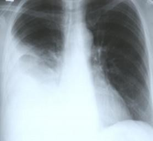
Figure 1: Showing right sided pleural effusion

Figure 2, 3 and 4. Showing huge cystic mass arising from right adnexal region with ascites
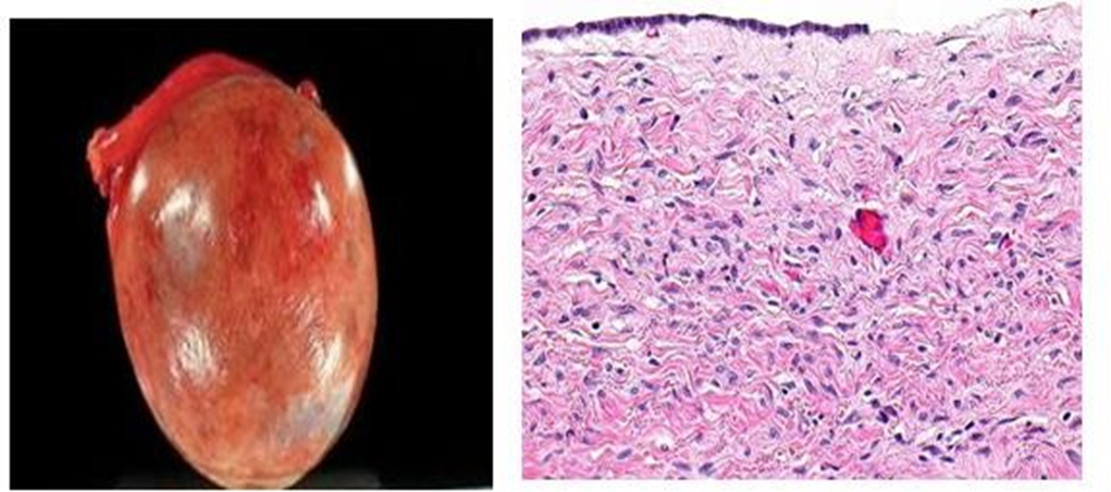
Figure 5 and 6: Serous Cystadenoma and histology of the tumor showing the cystic space is at the top of the image. Ovarian parenchyma is seen at the bottom right. H&E stain.
The combination of adnexal mass with pleural effusion and ascites in our case raised suspicion for Meigs’ syndrome. This syndrome is a rare disorder characterized by pleural effusion and ascites in patients with ovarian fibroma or fibroma-like tumors.[8] . It was first described in 1887 by Demons, and later in 1937 by Meigs, who arrived at the same findings about association of pleural effusion, ascites, and benign ovarian fibroma. Hence, this syndrome is also known as Demons-Meigs' syndrome.[9] . On the other hand, benign tumors of the ovary (other than fibromas) and ovarian malignancies, such as mature teratomas, mucinous cystadenoma, serous cystadenoma and struma ovarii, can also be associated with pleural effusions and ascites but they are categorized as pseudo-Meigs’ syndrome.[6] .
The pleurae are a structure of mesodermic origin that consists of two layers, denominated visceral and parietal layers. Both pleural layers unite in the base of the lung, leaving a space between each called the pleural cavity. A continuous process exists of filtration of liquid from the capillary vessels to the space subpleural, and give there to the cavity pleural. This process depends on the balance of hydrostatic and coloidosmotic pressures in both spaces, according to Starling's law, giving place to the presence of a minimal quantity of liquid in the physiologic virtual cavity. .[11] The pleural effusion in the cases of ovarian tumors, usually corresponds to an exudate because the liquid moves from the peritoneal cavity to the pleural cavity through diaphragmatic defects or lymphatic channels. It is generally located in the right and can be massive on occasions, with biochemical or cellular unspecific characteristics of the liquid.
Ovarian serous cystadenoma, also (less precisely) known as serous cystadenoma, is the most common ovarian neoplasm, representing 20% of ovarian neoplasms, and is benign [10]
It has a very superficial resemblance to the most common type of ovarian cancer (serous carcinoma of the ovary) under the microscope; however, .[1] it is virtually impossible to mix-up with its malignant counterpart (serous carcinoma), and [2] does not share genetic traits of indeterminate serous tumors, also called serous borderline tumors, that may transform into serous carcinoma [11].
Etiology of ascites has been explained by following mechanisms –
Vascular endothelial growth factor (VEGF) that raises capillary permeability is also reported to be associated with formation of pleural and peritoneal fluid. Ishiko et al. reported a significant difference between the VEGF levels in the pleural and peritoneal fluid before and after tumor removal in patients with Meigs’ or pseudo-meigs syndrome.[14] . The size of the pleural effusion is largely independent of the amount of ascites. The connection between the pelvic tumor and ascites is confirmed by the rapid resolution of abdominal and pleural fluid after removal of the tumor.
The most common presenting symptoms are dyspnea (due to pleural effusion), fatigue and weight loss and most of the patients initially referred to the general practitioner or chest physicians, .[15,16] .
The differential diagnosis for the presenting signs and symptoms include malignant ovarian tumor, other cancers including bowel and lung, nephrotic syndrome, congestive cardiac failure, liver cirrhosis and tuberculosis.
In spite of the valuable contribution of medical imaging techniques, the presumptive diagnosis of Meigs' or peuso-Meigs syndrome is made clinically. Upper abdominal ultrasound demonstrates ascites and should detect pleural effusions. Pelvic ultrasound demonstrates the presence of a well demarcated adnexal mass without increased vascularity. Chest x‐ray may be used to confirm the presence of a pleural effusion. Other imaging modalities like MRI or CT can be considered to exclude metastatic disease prior to treatment.
The cytological exam of the ascitic and pleural liquid in patients with ovarian tumors should be performed in order to differentiate between reactive processes and tumor spread. Although the detection of malignant cells is a marker of malignant disease and a sign of poor prognosis, the benign effusions don't affect neither the stage of the disease nor the prognosis of the patient.13 Review of reported cases of pseudo-Meigs syndrome shows that pleural fluid in these patients contains reactive mesothelial cells with no neoplastic cells25.Some authors emphasize that an ovarian mass with pleural and abdominal effusion doesn't always represent an advanced stage malignancy, not even in presence of high serum levels of CA-125 23,24.
CA-125 antigen is a tumor marker associated with ovarian carcinoma. Nevertheless, elevated levels of CA-125 have also been reported in the literature for Meigs or pseudo-meigs syndrome, although levels above 1,000 U/mL were rarely reported .[17,18] Lin et al. used immunohistochemical techniques to localize CA-125 expression, and they found that it is expressed by mesothelium rather than the fibroma .[19] . Case reviews have shown that higher levels of CA-125 are associated withhigher volume of ascites but size of tumor was not linearly correlated with CA-125 levels20.
The definitive diagnosis of Meigs' syndrome is usually postoperative with resolution of ascites and pleural effusions, and histological confirmation of the tumour. Serous cystadenomas are diagnosed by histomorphologic examination, by pathologists. Grossly, they are, usually, small unilocular cysts that contain clear, straw-coloured fluid. However, they may sometimes be multilocular. Microscopically, the cyst lining consists of a simple epithelium, whose cells may be either.[12] .
be columnar and tall and contain cilia, resembling normal tubal epithelium
be cuboidal and have no cilia, resembling ovarian surface epithelium
Medical care of patient with Meigs' or pseudo-Meigs syndrome involves paracentesis and thoracentesis for ascites and pleural effusion respectively. The treatment of choice is exploratory laparotomy with surgery and staging. Frozen section of ovarian mass is performed to confirm the benign nature of the mass. In women of reproductive age, unilateral salpingo oophorectomy is the treatment of choice, whereas in post‐menopausal women treatment is total abdominal hysterectomy with bilateral salpingo‐oophorectomy.
Removal of the tumor will ultimately result in resolution of ascites, pleural effusion, and normalization of CA-125 in Meigs’ and pseudo-Meigs’ syndrome .[5,22] . Life expectancy of patients after surgical removal of the tumor is the same as the general population.
In this case report, we presented a patient who had pleural effusions, ascites, normal CA-125 and right adnexal mass. An awareness of benign lesions of the pelvis with associated adverse features is important for both clinicians and their imaging partners to limited patient anxiety and direct appropriate treatment. Biochemical markers like CA-125 have limited diagnostic capabilities and their real value lies in cancer treatment surveillance.
Dear Editorial Team, Clinical Medical Reviews and Reports. My experience with the journal was highly positive. The peer-review process was rigorous, constructive, and completed in a timely manner. The reviewers provided valuable comments that helped improve the quality and clarity of our manuscript. The editorial office was professional, responsive, and supportive throughout all stages of the publication process. Communication was clear and efficient, and any questions were addressed promptly. Overall, I found the journal to maintain high scientific standards and an excellent publication workflow. I would be pleased to consider submitting future work to this journal. Best wishes from, Elena Popa.

It was my pleasure to submit my testimonial concerning the Reviewer Board of our Scientific Journal “Brain and Neurological Disorders”. The Reviewers focused on some modifications and their contribution was helpful. The ladies of our Editorial Office were also supported my efforts. It was my honor to have such a co-operation and I am looking forward for more collaboration.

Dear Grace Pierce, Editorial Coordinator of Journal of Clinical Research and Reports, Thank you for the speedy and efficient peer review process. I appreciate the fact that your peer reviewers do not take months to respond like with some other journals. I would also like to thank the editorial office for responding quickly to my questions. It is an excellent journal. I plan to submit more manuscripts in the future. Best wishes from, Robert W. McGee

Dear Grace Pierce, Editorial Coordinator of Journal of Clinical Research and Reports, Working with you and your team on our recent publication in JCRR has been a truly wonderful and enjoyable experience. The responses were prompt, and the reviewers were patient, constructive, and highly professional. One reviewer in particular gave me the feeling that a professor was carefully reading and commenting on my coursework, which was deeply touching. The entire process was straightforward and hassle‑free, with no tedious online forms to complete. I highly recommend this journal. Best wishes from, DR Aibing Rao, Head of R&D

I Appreciate the Opportunity to Share my Experience with the Journal of Clinical Research and Reports. The peer review process was timely and constructive, and the feedback provided helped improve the quality of our manuscript. The editorial office was professional, responsive, and supportive throughout the process, ensuring smooth communication and efficient handling of the submission. Overall, it was a positive experience collaborating with your team.

Dear Mercy Grace, Editorial Coordinator of Obstetrics Gynecology and Reproductive Sciences, We would like to express our gratitude for your help at all stages of publishing and editing the article. The editors of the magazine answer all the necessary questions and help at every stage. We will definitely continue to cooperate and publish other works in the Obstetrics Gynecology and Reproductive Sciences! Best wishes from, Alla Konstantinovna Politova,
