
Research Article | DOI: https://doi.org/10.31579/2578-8965/043
1 Department of Obstetrics and Gynecology, facultyof medicine, Ain Shams University, Cairo, Egypt.
2 Department of Obstetrics and Gynecology, faculty of medicine, Cairo University, Cairo, Egypt.
3 Department of Obstetrics and Gynecology. Faculty of Medicine Assiut University, Assuit, Egypt.
*Corresponding Author: Amr sobhy, Lecturer of Obstetrics and Gynecology, faculty of medicine, Ain Shams University, Cairo, Egypt.
Citation: Amr Sobhy, Suzy Abdel Aziz, Sherif Mohammed Abdel-Mageed Badran, Doaa Fouad, Hanan Shehata, et al., (2020). A Prospective Cohort study on Ovarian Vascular and Hormonal Changes in Patients with Polycystic Ovarian Syndrome after Laparoscopic Ovarian Drilling. J. Obstetrics Gynecology and Reproductive Sciences, 4(4) DOI:10.31579/2578-8965/043
Copyright: © 2020 Amr Sobhy. This is an open-access article distributed under the terms of The Creative Commons Attribution License, which permits unrestricted use, distribution, and reproduction in any medium, provided the original author and source are credited.
Received: 02 November 2020 | Accepted: 09 November 2020 | Published: 18 November 2020
Keywords: PCOS; laparoscopic ovarian drilling; 3D- ultrasound
Background:
Laparoscopic ovarian drilling (LOD) is a well-known modality for treatment of anovulatory PCOS.
Aim:
The aim of this study is to determine the effect of laparoscopic ovarian drilling on ovarian reserve in terms of measuring FSH, E2, AFC and (3D) power Doppler indices (Vascularization index, Flow index and Vascularization flow index) of ovarian stromal blood flow as ovarian reserve markers.
Design:
This prospective cohort study was conducted in 2 hospitals in Saudi Arabia, from period of January 2019 to December 2019.
Methods:
Forty eight patients were including in the study, all were candidates for LOD, preoperative D2 or Day 3 serum FSH, LH And E2 were measured and 3D US was performed to measure ovarian volume, AFC and ovarian stromal blood flow indices (VI, FI and VFI). Laparoscopic ovarian drilling was done postmenstrual then post laparoscopy by 3-month the hormonal profile (Day 2-3 FSH,LH and E2) were measured and 3D US was repeated for all patients.
Results:
Serum E2 and FSH level was compared before and 3 months after laparoscopic ovarian drilling and the results showed that there was no statistically significant difference after LOD (p>0.05). In the current study, there was a statistically significant difference in each of serum LH level, AFC, ovarian volume and ovarian stromal blood flow indices before and 3 months after laparoscopic ovarian drilling (p<0.001).
Conclusion:
Bilateral laparoscopic ovarian drilling in PCOS patients decrease serum level of LH, ovarian volume, AFC and 3D power Doppler indices of stromal blood flow of ovaries of PCOS patients after LOD.
Polycystic ovary syndrome (PCOS) is the most common endocrine, metabolic disorder in the reproductive age with a genetic origin. PCOS is responsible for 75% of female infertility due to anovulation. Although the pathogenesis of polycystic ovary disorder is complex and not fully understood, some studies have shown that hyperandrogenism and hyperinsulinemia are main contributors to this disorder [1].
The most relevant markers of ovarian reserve are serum FSH, AMH and antral follicle count (AFC) measured by transvaginal ultrasound. AMH is produced pre antral and antral follicles measuring 6–8 mm diameter then secreted in blood. AFC is counted by transvaginal ultrasound (antral follicles of 2–10 mm present in both ovaries). Three-dimensional ultrasound can accurately measure the ovarian volume, number and size of antral follicles, while the Three-Dimensional power doppler can quantify ovarian blood flow with its indices (vascularization index VI, flow index FI and vascularization flow index VFI) [2].
Laparoscopic ovarian drilling (LOD) has been invented several decades ago and still used as an effective treatment of polycystic ovarian syndrome (PCOS) in comparison to gonadotropin induction. However, it has its own side effects on decreasing the ovarian reserve [3]. Laparoscopic ovarian drilling used as an alternative to gonadotrophin clomiphene-resistant PCOS and it increases the ovulation rate and the cumulative pregnancy rate [4]. Laparoscopic ovarian drilling (LOD) causes a transient increase then significant decrease in ovarian volume as measured by 3D ultrasound LOD also affects the ovarian stromal flow after a period of short-term follow-up [5].
Aim of this study is to determine the effect of laparoscopic ovarian drilling on hormonal changes in terms of measuring FSH, LH, E2, AFC and vascular changes as measured by (3D) power Doppler indices (VI, FI and VFI)
Study design:
This prospective cohort study was conducted on 48 patients at 2 different hospitals in Saudi Arabia from the period of January 2019 to December 2019
Participants:
Participants were included in the study according to the following criteria:
Inclusion criteria: Women aged between 18 and less than 35 years, PCOS was diagnosed according to the (Rotterdam Criteria, 2003) on presence of any 2 of the following parameters: oligomenorrhea and/ or anovulation, clinical and/or biochemical signs of hyperandrogenism and finally polycystic ovaries by transvaginal ultrasound that should have at least one of the following: either 12 or more follicles measuring 2-9 mm in diameter, or increased ovarian volume ( greater than10cm3).They were Clomiphene citrate resistant (CC-resistant).
The Exclusion criteria were women aged below 18 years or more than 35 years, women with single ovary, previous ovarian cystectomy, thyroid disease, diabetes or any other endocrinopathy.
The procedure was explained to all the participants in this study, written consent was taken from every participant. All the participants were subjected to the following: full history and examination especially for signs of hyperandrogenism. Day 2 of menses before Laparoscopy blood samples were taken to assess hormonal profile (FSH, LH and serum E2). Transvaginal 3D US was done to calculate ovarian volume, mean antral follicle number in both ovaries (measuring 2-9mm) and three indices quantifying the power Doppler signal were determined namely, vascularization index (VI), flow index (FI) and vascularization flow index (VFI).
Bilateral laparoscopic ovarian drilling was carried out postmenstrual. In Laparoscopy; bilateral ovarian bilateral ovarian drilling was done using monopolar electrocautery needle perpendicular to ovarian surface aided by a short duration of cutting current of 30 W for 4-6 seconds in 4 punctures. All operations were done by same level and experienced consultants. Three months later after LOD, hormonal profile was repeated for all patients to measure (LH, FSH and Serum E2) and 3D US (AFC, Ovarian volume and doppler indices). The 3D US were performed by the same investigators and were both of same level and experience.
Sample size justification:
Sample size was calculated using STATA® version 11 programs, setting the type-1 error (α) at 0.05 and the power (1-β) at 0.9. Results from a previous study (Salem et al., 2017) Calculation according to their results produced a minimal sample size of 43 cases. However, we included 48 cases to compensate for drop out cases
Statistical analysis
Statistical analysis was done using Statistical package for Social Science (IBM Corp. Released 2017. IBM SPSS Statistics for Windows, Version 25.0. Armonk, NY: IBM Corp). Shapiro wilk’s test was used to evaluate normality of Quantitative variables which was presented as mean and standard deviation, Paired t-test was used to assess the statistical significance of the difference between two means measured twice for the same study group. P less than 0.05 is considered statistically significant.
As regard the Comparison of FSH,LH and E2 levels before and after laparoscopic ovarian drilling (LOD); Table 1 shows that there was there was no statistically significant difference between patients before and after LOD regarding the serum FSH and serum E2 (p greater than0.05). Statistically, there was a statistically significant decrease between patients before and after LOD regarding the serum LH (p less than0.001).
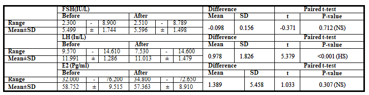
Table 1: Comparison of hormonelevels before and after laparoscopic ovarian drilling (LOD).
The drilling of ovaries causes statistically significant decrease in all sonographic studied parameters as shown in Table 2 (ovarian volume, AFC). Table 3 showed statistically significant decrease in the vascularization index, flow index and vascularization flow before and after LOD
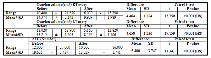
Table 2: Comparison of ovarian volume of the right and left ovary and antral follicular count (AFC) before and after laparoscopic ovarian drilling (LOD
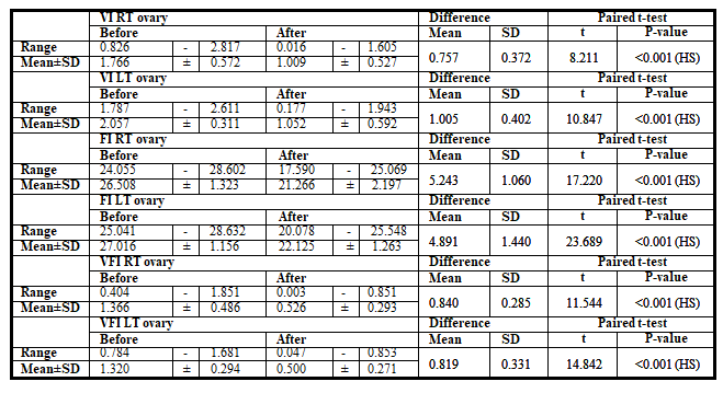
Table 3: Comparison of Vascularization index (VI), flow index (FI) and vascularization flow index (VFI) of the right and left ovary before and after laparoscopic ovarian drilling (LOD)
Our results interpretation and its comparison to other studies
The present study was done on 48 women with PCOS fulfilling the (Rotterdam Criteria, 2003) to whom bilateral laparoscopic ovarian drilling was carried out postmenstrual. Serum (FSH, LH and E2 levels), AFC, ovarian volume and ovarian stromal blood flow indices were compared before and after drilling by 3 months.
The results of the present study agree with the results of Amer et al.,2017 who had a prospective controlled clinical study that included anovulatory women with CC-resistant PCOS who underwent LOD and showed that there was no statistically significant difference between patients before and after
LOD regarding the serum FSH (p greater than0.05) [7]. However, the results of the current study disagreed with the results of (Rezk et al., 2016) who had a study on twenty-one PCOS women who underwent LOD and day-3 serum FSH levels were significantly higher after drilling (p less than0.001) [8].
In the current study as regard serum LH level, the results showed that there was statistically significant decrease after drilling (p less than0.001). These results agreed with the results of (Amer et al., 2017) who had a prospective clinical study included anovulatory women with CC-resistant PCOS who underwent LOD and showed that there was statistically significant decrease between patients before and after LOD regarding the serum LH (p less than0.001) [7]. Also,
they agreed with study of study of (Rezk et al 2016.) where study showed that LH decreased significantly in the 1st and 6th months after LOD [8].
In the present study as regard serum E2 level, the results showed that there was no statistically significant difference (p greater than0.05) after LOD. These results disagreed with the results of (Rezk et al., 2016) who studied women with CC-resistant PCOS who underwent LOD and showed that there was statistically significant increase in serum E2 levels after LOD (p less than0.001) [8].
In the present study, mean antral follicle count and ovarian volume of both ovaries were compared before and 3 months after laparoscopic ovarian drilling and the results showed that there was statistically significant decrease after LOD (p less than0.001) regarding both parameters.
In the current study, ovarian indices (VI, FI and VFI) in women with PCOS were measured using 3Dpower Doppler ultrasound before and after laparoscopic ovarian drilling by 3 months and the results revealed a statistically significant decrease in ovarian indices after LOD (p less than0.001).
The current study agreed with study of Kamal et al., (2018) which was carried on 80 Patients Pre- and post-LOD ovarian reserve parameters (ovarian volume: OV, and antral follicle count: AFC) and ovarian stromal blood flow indices (Vascularization index: VI, flow index: FI, and vascularization flow index: VFI) were measured to explore the effect of LOD and to find out the correlation between different clinical, hormonal, and ultrasonic variables. There was a highly significant reduction in OV, AFC and vascular indices (VI, FI and VFI) of the right and left ovaries (p less than .05). LOD significantly reduced ovarian reserve parameters (OV and AFC) and ovarian stromal blood flow indices (VI, FI and VFI) [9].
Unfortunately, the results of the current study disagreed with the results of (Alia, 2014) who studied 53 women with polycystic ovary syndrome who underwent LOD. Serum E2 and FSH levels and ovarian stromal blood flow indices (VI, FI and VFI) were measured and compared before and after LOD. The results of the study showed a statistically significant increase between patients before and after LOD (p less than0.001) regarding serum E2 and FSH levels and ovarian stromal blood flow indices (VI, FI and VFI) [9].
The cause that the results of the current study were different from the results of the above-mentioned study may be attributed to the technique of how laparoscopic ovarian drilling was performed (number, depth of punctures, type of electrocautery needle, time of application of needle to ovarian surface and the power of setting electrocautery). In the current study, bilateral LOD was done by using monopolar electrocautery needle which was applied perpendicular to the ovarian surface aided by a short duration of cutting current 30 W for 4-6 seconds in 4 punctures.
Another issue that may explain why the results of the current study (regarding ovarian indices) were different from previous studies is the timing of performing the 3D power Doppler ultrasound. In the current study, the 3D power Doppler indices (VI, FI and VFI) were measured in second or the third day of the menstruation before and after LOD. In the other study, the 3D power Doppler indices (VI, FI and VFI) were measured in day 11 or 12 of the menstrual cycle before and after LOD [8].
Strength and weakness of study
Strength of our study is the correlation of hormonal and vascular changes that occurred after LOD, limitation of our study is small number of participants
Clinical implications of Study
Although Laparoscopic ovarian drilling is an alternative treatment to induction of ovulation by gonadotrophins in patient with clomiphene citrate resistance, but it has its effect on ovarian reserve so we suggest to do hormonal profile and 3D power doppler pre and post laparoscopy to see its effect on hormonal and vascular changes.
Recommendations for further studies
Further studies are needed to see effect of other parameters as AMH and effect of LOD
Bilateral laparoscopic ovarian drilling in PCOS patients decrease serum level of LH, ovarian volume, AFC and 3D power Doppler indices of stromal blood flow of ovaries of PCOS patients after LOD.
Ethics approval:
Study approved by Ethical Committee of 2 participated hospitals
Availability and data material:
The data sets used and/or analyzed during the current study are available from the corresponding author on reasonable request.
Competing interests:
The authors report there are no competing interests to declare
Funding:
This study received no financial support.
Dear Editorial Team, Clinical Medical Reviews and Reports. My experience with the journal was highly positive. The peer-review process was rigorous, constructive, and completed in a timely manner. The reviewers provided valuable comments that helped improve the quality and clarity of our manuscript. The editorial office was professional, responsive, and supportive throughout all stages of the publication process. Communication was clear and efficient, and any questions were addressed promptly. Overall, I found the journal to maintain high scientific standards and an excellent publication workflow. I would be pleased to consider submitting future work to this journal. Best wishes from, Elena Popa.

It was my pleasure to submit my testimonial concerning the Reviewer Board of our Scientific Journal “Brain and Neurological Disorders”. The Reviewers focused on some modifications and their contribution was helpful. The ladies of our Editorial Office were also supported my efforts. It was my honor to have such a co-operation and I am looking forward for more collaboration.

Dear Grace Pierce, Editorial Coordinator of Journal of Clinical Research and Reports, Thank you for the speedy and efficient peer review process. I appreciate the fact that your peer reviewers do not take months to respond like with some other journals. I would also like to thank the editorial office for responding quickly to my questions. It is an excellent journal. I plan to submit more manuscripts in the future. Best wishes from, Robert W. McGee

Dear Grace Pierce, Editorial Coordinator of Journal of Clinical Research and Reports, Working with you and your team on our recent publication in JCRR has been a truly wonderful and enjoyable experience. The responses were prompt, and the reviewers were patient, constructive, and highly professional. One reviewer in particular gave me the feeling that a professor was carefully reading and commenting on my coursework, which was deeply touching. The entire process was straightforward and hassle‑free, with no tedious online forms to complete. I highly recommend this journal. Best wishes from, DR Aibing Rao, Head of R&D

I Appreciate the Opportunity to Share my Experience with the Journal of Clinical Research and Reports. The peer review process was timely and constructive, and the feedback provided helped improve the quality of our manuscript. The editorial office was professional, responsive, and supportive throughout the process, ensuring smooth communication and efficient handling of the submission. Overall, it was a positive experience collaborating with your team.

Dear Mercy Grace, Editorial Coordinator of Obstetrics Gynecology and Reproductive Sciences, We would like to express our gratitude for your help at all stages of publishing and editing the article. The editors of the magazine answer all the necessary questions and help at every stage. We will definitely continue to cooperate and publish other works in the Obstetrics Gynecology and Reproductive Sciences! Best wishes from, Alla Konstantinovna Politova,
