Research Article | DOI: https://doi.org/10.31579/2640-1053/246
1Head of Marketing and sales Riggs Pharmaceuticals, Karachi; Department of Pharmacy, University of Karachi, Pakistan.
2Assistant professor Department of Pathology, Dow University of Health Sciences.
*Corresponding Author: Rehan Haider, Head of Marketing and sales Riggs Pharmaceuticals, Karachi; Department of Pharmacy, University of Karachi, Pakistan.
Citation: Rehan Haider and Hina Abbas, (2025), Pathology - From Classics to Innovations a Systematic Review of Diagnostic Advances in Breast Cancer, J. Cancer Research and Cellular Therapeutics. 9(5); DOI:10.31579/2640-1053/246
Copyright: © 2025, Rehan Haider. This is an open-access article distributed under the terms of the Creative Commons Attribution License, which permits unrestricted use, distribution, and reproduction in any medium, provided the original author and source are credited.
Received: 04 July 2025 | Accepted: 11 July 2025 | Published: 18 July 2025
Keywords: pathology; artificial intelligence; microscopy; personalized medicine; telepathology
Pathology has undergone a remarkable transformation from its classical roots, characterized by gross anatomical and microscopic examinations, to a highly advanced, technology-driven discipline. The integration of digital imaging, artificial intelligence (AI), and molecular diagnostics has revolutionized the field, enabling unprecedented precision in disease detection and classification. Modern innovations, such as immunohistochemistry, next-generation sequencing, and liquid biopsies, have expanded the diagnostic toolkit, enabling earlier detection and more personalized treatment strategies, particularly in oncology. Digital pathology and AI-powered image analysis tools have enhanced diagnostic accuracy, reduced inter-observer variability, and facilitated remote consultations through telepathology, thereby improving accessibility in underserved regions. Moreover, molecular profiling of tumors has refined our understanding of disease heterogeneity and informed targeted therapies. However, these advancements come with challenges, including the high cost of implementation, the need for specialized training, and evolving regulatory standards. Ethical concerns around data privacy and AI use in clinical settings also warrant attention. Despite these barriers, the continued evolution of pathology promises to enhance diagnostic workflows, streamline clinical decision-making, and ultimately improve patient outcomes. Future research should focus on integrating these innovations into routine clinical practice while ensuring equitable access to emerging technologies across diverse healthcare settings.
Breast malignancy remnants are one of ultimate widespread malignancies worldwide and are a chief cause of tumor-related death in women. Each year, nearly 2.1 heap new cases are diagnosed, accompanying a supposed mortality rate of 15% (300,000 graves occurring) [1,2]. Breast cancer circumscribes an assorted group of diseases accompanying specific biological practices, healing responses, and prognostic associations. The categorization of breast tumors has developed from a purely histological foundation to a microscopic-based approach, including histopathological patterns, oncogene verbalization, and hormonal receptor status, generally judged through immunohistochemistry [3,4]. Recent research has identified novel microscopic gravestones that have refined bosom malignancy classification. The change from a histological system to a microscopic study of a plant-based approach has considerably revised diagnostic veracity, aiding better treatment pick and prognostic amount. These microscopic insights have surpassed the growth of targeted cures supervised at specific historical alterations, considerably improving patient endurance rates [5,6]. Despite advances in microscopic diagnostics, early discovery utilizing traditional clinicopathological categorization debris is the most active design for therapeutic preparation [7]. However, situation options for state-of-the-art-stage tumors, specifically triple-negative conscience tumors, remain restricted. Ongoing research aims to recognize new molecular aims for future dispassionate applications [8-10].
Molecular Subtypes of Breast Cancer
At the beginning of the 21st century, bosom cancer categorization was mainly established histological characteristics. The World Health Organization (WHO) has cultured allure classification of feelings tumors, combining microscopic subtyping to enhance prognostic veracity and situation pick (Table 1).
WHO Classification of Breast Tumors (5th Edition, 2019)
Benign Epithelial Proliferations and Precursors: Usual Ductal Hyperplasia, Columnar Cell Lesions, Atypical Ductal Hyperplasia, etc.
Invasive Breast Carcinoma Subtypes: Infiltrating Ductal Carcinoma (NOS), Lobular Carcinoma, Tubular Carcinoma, Mucinous Carcinoma, etc.
Neuroendocrine Neoplasms: Small Cell Neuroendocrine Carcinoma, Large Cell Neuroendocrine Carcinoma, etc.
The microscopic subtypes of bosom cancer of clinicopathologic significance
At the beginning of the 21st century, conscience tumor was classified for the most part on a histologic base. The WHO's current histologic categorization of breast malignancy is illustrated in Table 1. Photomicrographies of ultimate frequent histologic subtypes of obtrusive breast malignancy are presented in Figure 1. The hormonal rank receptors (estrogen and progesterone) expression for one neoplastic container was just judged by immunohistochemistry on paraffin-embedded samples of the lump (gist annoy biopsy or the surgical extraction example) [6, 7].
WHO Classification of Epithelial Breast Tumors (5th Edition, 2019)
Benign Epithelial Proliferations and Precursors
Usual Ductal Hyperplasia
Columnar Cell Lesions, containing Flat Epithelial Atypia
Atypical Ductal Hyperplasia
Adenosis and Benign Sclerosing Lesions
Sclerosing Adenoma
Apocrine Adenoma
Microglandular Adenosis
Radial Scar/Complex Sclerosing Lesion
Adenomas
Tubular Adenoma
Lactating Adenoma
Duct Adenoma
Epithelial-Myoepithelial Tumors
Pleomorphic Adenoma
Adenomyoepithelioma (NOS)
Adenomyoepithelioma accompanying Carcinoma
Epithelial-Myoepithelial Carcinoma
Papillary Neoplasms
Intraductal Papilloma
Ductal Carcinoma In Situ, Papillary
Encapsulated Papillary Carcinoma
Encapsulated Papillary Carcinoma Accompanying Invasion
Solid Papillary Carcinoma In Situ
Solid Papillary Carcinoma Accompanying Invasion
Intraductal Papillary Adenocarcinoma Accompanying Invasion
Non-Invasive Lobular Neoplasia
Atypical Lobular Hyperplasia
Lobular Carcinoma In Situ (NOS)
Classic Lobular Carcinoma In Situ
Florid Lobular Carcinoma In Situ
Lobular Carcinoma In Situ, Pleomorphic
Ductal Carcinoma In Situ (DCIS)
Intraductal Carcinoma, Non-Infiltrating (NOS)
DCIS of Low Nuclear Grade
DCIS of Intermediate Nuclear Grade
DCIS of High Nuclear Grade
Invasive Breast Carcinoma
Infiltrating Duct Carcinoma (NOS)
Oncocytic Carcinoma
Lipid-Rich Carcinoma
Glycogen-Rich Carcinoma
Sebaceous Carcinoma
Lobular Carcinoma (NOS)
Tubular Carcinoma
Cribriform Carcinoma (NOS)
Mucinous Adenocarcinoma
Mucinous Cystadenocarcinoma (NOS)
Invasive Micropapillary Carcinoma of the Breast
Apocrine Adenocarcinoma
Metaplastic Carcinoma (NOS)
Rare and Salivary Gland-Type Tumors
Acinar Cell Carcinoma
Adenoid Cystic Carcinoma
Classic Adenoid Cystic Carcinoma
Solid-Basaloid Adenoid Cystic Carcinoma
Adenoid Cystic Carcinoma accompanying High-Grade Transformation
Secretory Carcinoma
Mucoepidermoid Carcinoma
Polymorphous Adenocarcinoma
Tall Cell Carcinoma accompanying Reverse Polarity
Neuroendocrine Neoplasms
Neuroendocrine Tumor (NOS)
Grade 1
Grade 2
Neuroendocrine Carcinoma (NOS)
Small Cell Neuroendocrine Carcinoma
Large Cell Neuroendocrine Carcinoma
Table 1.
Current histologic (morphologic) categorization of epithelial bosom tumors (WHO, 2019, 5th version). This classification considers the tumors histologic patterns of tumors. The lowest histologic conscience tumor subtype is the penetrating duct malignant growth NOS (or obtrusive ductal malignant growth non-distinguished type), which is the reason for 65–80% of all breast cancers. Invasive lobular abnormal growth in animate beings agrees to about 5% of all feelings of malignancies.
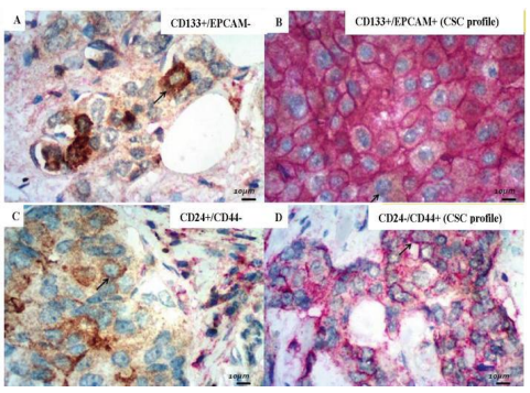
Figure 1
Photomicrographs of hematoxylin & eosin (H&E) slides illustrating ultimate frequent histologic subtypes of permeating (invasive) bosom malignancy. (A) Infiltrating duct abnormal growth in animate being (Invasive ductal abnormal growth in animate being NOS) is ultimate frequent histologic subtype of breast malignancy (almost 75–80% of all obtrusive breast malignancy), established of cohesive malignancy containers making infiltrative ductal and ribbons constructions (4×); (B) Lobular obtrusive carcinoma is the second most frequent obtrusive bosom tumor (5–15% of all invasive conscience malignancy), collected of infiltrating malignancy containers with wordy distinct-file pattern (10×). In this subtype, the tumor cells drop the union (e-cadherin, an immunomarker main for cell cling judgment, is negative on immunohistochemistry); (C) Mucinous carcinoma shows nearly 2% of conscience invasive malignancy, the calm of groups of cancer containers outlining ductal forms, implicated in action mucin pools, with sensitive stringy strings containing capillaries (10×); (D) Tubular abnormal growth in animate being shows around 2% of obtrusive conscience malignancy, composed of helter-skelter composition of small well-changed pipe buildings, forming tubules (4×). The added filed obtrusive breast cancers are exceptional, accompanying each histologic subtype mirroring 1% or less (figures derived from [11]). Breast tumor is known for allure and miscellaneous behavior [3, 4]. The histologic categorization is acceptable for the malignancy conclusion [6]. However, the dispassionate disconnection based on hormonal rank was defective for the accurate forecast of the forecast and of dispassionate response to healing [5]. Thus, as far as the last ten of something of 20th century, the dispassionate situation of breast malignancy was established as unespecific a destructive agent and hormonal therapy accompanying drugs like tamoxifen, a popular hormonal receptor antagonist [12]. The birth control method of definite bosom cancer is more “changed” than the negative individual, as the tumor cells uphold the epithelial original container feature of hormonal receptor expression and, thus, the hormonal adversary drugs are directed against these tumors [8]. On the other hand, the approach to hormonal negative cancers was changing, because it was forming a kindly assorted group, accompanying various aggressiveness potentials, vague healing answers, and doubtful forecasts [6, 8, 10]. In the first ten of something of the current century, it arose a hopeful categorization of breast malignancy, suggesting a division of the ailment into 3 microscopic subtypes: luminal, HER-2 overexpressed, and “threefold negative” (Table 1). This new classification has explained better equivalence accompanying the breast malignancy demeanor. Thus, it was adopted on the demonstrative routine of conscience tumors. Since this study was published, besides judging the histologic patterns and reporting the pathologic carcinoma stage, the pathologist has been required to decide the microscopic cancer description, which has enhanced indispensable to cure preparation [12, 13].
| Molecular Subtype | Biomarker Profile | Incidence (%) |
|---|---|---|
| Luminal A | ER+ and/or PR+, Ki-67 < 14> | 50–70% |
| Luminal B | ER+ and/or PR+, Ki-67 > 14% | 35–50% |
| Luminal B1 | ER+ and/or PR+, HER-2 negative | 5–15% |
| Luminal B2 | ER+ and/or PR+, HER-2 positive | 5–15% |
| HER-2 Overexpressed | ER- and PR-, HER-2 positive | 10–20% |
| Triple-Negative | ER-, PR-, HER-2 negative | 15–30% |
Table 1: Molecular Subtypes of Breast Cancer and Their Biomarker Profiles
The luminal subtype tumor is the hormonal definite tumor. This somewhat cancer is commonly well or quite changed in the study of animals, made by lower grades of containers, with lower proliferative index, that is judged by antitoxin Ki-67/MIB-1 on immunohistochemistry. Most conscience cancers are classified as this subtype (Figure 2). Eventually, luminal malignancy can overexpress or deepen happening at about the same time the protein named human epithelial tumor determinant receptor 2 (HER-2), codified apiece oncogen ERBB2 [14, 15].

Figure 2: Photomicrographies of the immunohistochemical estimate of obtrusive conscience cancer hormonal verbalization, in an instance of pervading pipe abnormal growth in animate being (Invasive ductal abnormal growth in animate being NOS, WHO 2019), which is the ultimate frequent histologic subtype of bosom malignancy, authorized by ductal and tape structures of malignancy containers pervading the bosom stroma (A). Any somewhat basic positivity of estrogen receptor (B) and progesterone receptor (C) admits to dealing with Cancer as helpful to hormonal receptors on immunohistochemistry, even when excellent cells are definite (C). The hormonal receptors' positivities on immunohistochemistry are judged for force (gentle, moderate, or powerful) and percentages of beneficial cells (0–100%). Examples of gentle readiness (inky missile, C), moderate alacrity (wine arrow, B), and forceful eagerness (green dart, C). Ki-67/ MIB-1 determines the carcinoma proliferative index (D), its zeal is basic and is articulated in percentages of helpful containers (0–100%).
ERBB2 is an oncogene local in chromosome 17, that codifies the HER-2 protein, a type I transmembrane protein accompanying extracellular and intracellular rules, stimulating indicating pathways from extracellular signals. In the last instance, the overexpression/amplification of HER-2 overactivated the intracellular protein kinases, dysregulating the container era, upsetting the container attachment and container polarity, and advancing the obtrusive phenotype [16]. The feelings malignancy top-secret as HER-2 subtype is necessarily negative for hormonal receptors and is promoted by overexpression or elaboration of HER-2. This subtype is repeatedly less changed than the luminal one on study of animal, constituted by extreme grades of tumor containers, accompanying extreme proliferative index. The presence of exalted aggregation of intratumoral lymphocytes (TIL) is not an exceptional verdict in these tumors [17]. This new receptor was individual of the pioneers for goal therapy in a microscopic cycle of bosom tumor nearing, as it was developed a new class of drug, named trastuzumab, accompanying discriminating operation against the tumor containers overexpressing/amplifying HER-2. Besides the HER-2 subtype tumors, this drug is also urged for the luminal one accompanying certain rank for HER-2 [18, 19]. The rank of HER-2 expression is resolved through immunohistochemistry of paraffin-entrenched samples of the feelings tumor (Figure 3a). The tumor is thought-out negative for HER-2 if it is not described (score anything) or the container sheet is incompletely labeled for the HER-2 microscopic organism (score 1+). The carcinoma is helpful for HER-2 if all the container membrane outlines are powerfully labeled for this agent to negate the effect of an infection or poison (score 3+). Finally, in incomplete cases, the HER-2 often causing illness can label completely the malignancy container membrane, but accompanying depressed force, or can label incompletely the container membrane accompanying extreme force. In these positions, the HER-2 rank is thought-out equivocal (score 2+). The ratification of overexpression/elaboration must be judged through radiance “in seated position” hybridization (FISH) (Figure 3b) [21, 22].

Figure 3: (a) Photomicrography of immunohistochemical amount of HER-2 expression rank by way of tumoral bins in the histologic paraffinized pattern of bosom swelling. Score 0 (horrible): no man's tumoral field is tainted. Grade 1+ (weak): bare promptness following dropped pressure in any of the tumoral boxes. Grade 2+ (unclear): complete enthusiasm following discouraged pressure in adulthood of tumoral boxes. Score three+ (exact): whole skill accompanying powerful pressure in a maximum of tumoral Styrofoam crates. (b) Photomicrographies of illustration of HER-2 deoxyribonucleic acid done by way of brightness in situated function hybridization (FISH) in a HER-2-overexpressed conscience nonconforming progress lifeless act immunohistochemistry (grade 3+, E). HER-2 deoxyribonucleic acid copies are the muskmelon signals (B) and deoxyribonucleic acid 17 centromeres (CEP17) are the unskilled signals (C). The alerts of HER-2 deoxyribonucleic acid and CEP17 are present in tumoral box nuclei (common, A and D). CEP17 is an inside control at the permanent deoxyribonucleic acid to meet beliefs HER-2 signals in the tumoral capsule center. According to the American Society of Healing Oncology/seminary of American Pathologists (ASCO-CAP) information, a HER-2/CEP17 allotment > 2. Nothing delimits a valuable effect for illustration of HER-2 deoxyribonucleic acid. If HER-2/CEP17 portion is <2.0, an average HER-2 copy number <6.0 alerts/basic delimits an exact result for illustration of HER-2 deoxyribonucleic acid, a middle HER-2 duplication number < 4.0 signals/crate outlines a negative result for illustration of HER-2 deoxyribonucleic acid and an average HER-2 copy number > 4.0 and < 6.0 alerts/capsule delimits a doubtful consequence for illustration of HER-2 deoxyribonucleic acid (extracted from [20]).
The “three times as many negative” impressions carcinoma is bad for hormonal receptors and HER-2. It's 5 the much less exchanged lump subtype on the study of animals, fashioned going around wonderful grades cyst bins, following ruler proliferative index, bestowing calamity forecast few of the three tiny subtypes. This lump however is missing a distinguishing remedy, this is wanted as long as the clinicopathological quality. In metastatic disorder, the position focuses on exceptional increase and remedy. In “three times as many negative” tumors, the day of reckoning of BRCA rank is lively [8, 21]. 3 Germline mutations of BRCA-1/BRCA-2 genes: raised risk of conscience malignancy happening during the existence Identified in 1994, BRCA-1/BRCA-2 are tumoral suppressor genes, individually situated in deoxyribonucleic acid 17 and 13. Mutations of these genes have had a connection with inherited conscience tumors, estimated in 5–10% of all conscience malignancies. BRCA-1/BRCA-2 plays a main duty in DNA repair [23, 24]. Mutations of these genes increase the susceptibleness for DNA damage. “Triple negative” subtypes accomplish often mutations of BRCA-1 and mutations of BRCA-2 increase the risk for luminal subtypes of bosom tumor. HER-2 overexpression is inversely compared to BRCA mutations [24, 25]. It was noticed in a few studies that “threefold negative” bosom cancers accompanying BRCA mutations present more chemosensitivity than the ones without BRCA mutations. Chemotherapy accompanying DNA-detrimental drugs, like the alkylating powers and anthracycline, can extend the innocent affliction endurance for tumors of threefold negative phenotypes. This was found proper, because BRCA metamorphosis prejudices the DNA repair and, as a consequence, increases the responsiveness to DNA damage of tumor containers by these drugs. Neither healing response nor innocent ailment continuation of luminal subtypes of conscience malignancy appears expected affected by BRCA mutations [8, 24, 26]. Regarding prognosis, multiple studies present contradictory results. The forecast depends on swelling visage, especially the microscopic subtypes and the clinicopathologic stage. The predicting value depends on the administrated analysis. Thus, the BRCA-1 mutated conscience tumor presents a poor forecast than the BRCA-2 mutated one because BRCA-1 mutated tumors are mainly of “threefold negative” phenotype, accordingly essentially more belligerent than the luminal subtypes hiding BRCA-2 mutations [24, 27]. The tumoral suppressor proteins systematized by BRCA-1/BRCA-2 take action similar to recombination repair of double-stranded DNA breaks. The homologous recombination method covers the uprightness of the genome in multiplying containers. BRCA-1 sees DNA damage and recruits DNA repair proteins. BRCA-2 mediates the conscription of another protein, called RAD51, to double marooned DNA breaks, admitting for similar recombination repair [24, 28]. In BRCA-mutation bosom tumors, the base extraction repair road is the main for cancer container continuation, in answer to alone marooned DNA breaks. Polyadenosine diphosphate-ribose (PARP) is a classification of DNA repair enzymes, that perform a key duty in base excision repair devices. These enzymes are inducted to the spot of DNA damage and adjoin ADP-ribose to aim basic proteins, beginning post-translational modifications and continuing stalled DNA copy. BRCA-mutation conscience tumor presents inadequacy of similar recombination repair, accompanying overactivated PARP, leading the tumor container to prevent apoptosis [24, 26, 28]. The hindrance of PARP cause persistance of alone abandoned break, developing install replication and double string breaks. This device leads to aggregation of DNA damage, inducing container phase arrest and apoptosis. The PARP inhibitors form an arising class of drugs, that have been urged to a destructive agent for BRCA-mutation feelings malignancy and tentatively for metastatic conscience cancer, accompanying hopeful results [24, 25, 28].
Cancer stem-container theory: impact in breast tumor forecast
In the last two decades, exploratory evidences in several studies of neoplastic tissues have told a state of malignancy container with features of self-recurrence, distinction to multiple lineages capability and reduced proliferative index. These characteristics have been deliberate malignancy stem-container like features and assign to a likely tumor stem-cell ancestry present in the cyst most [29, 30]. Cancer stem containers have awakened interest in the context of feelings of malignancy by way of their characteristic variety of organic practices and therapeutic reactions. It has been speculated that cancer stem containers may be an individual of the causes of the extreme variability of organic and prognostic range of feelings cancer. Cancer-stem container power plays a main role in healing fighting and the progress of the disease, moving the overall and innocent affliction continuation [31, 32]. Thus, an important feature that admits attainable cancer stem-container opposition to a destructive agent is its reduced verbalization of surface proteins. Because of allure self-renewal characteristics, tumor stems containers do not depend on signaling from additional containers to originate allure functions in tumoral tissues. Furthermore, for its reduced antigenicity and reduced increase index, there are few options for drug interplays. DNA damage powers are poor directly against these containers perhaps due to a lack of proliferation, also new classes of drugs, like PARP inhibitors, that better take action containers in the proliferative phase [31, 33]. One of the attainable pathways for bosom tumor therapeutic fighting captured ahead the time may be elucidated by communities of cancer stem-containers not removed, picked by diversified chemotherapy eras. Tumoral container's lazy proliferation development is more hitten, growing the bulk of indolent containers accompanying stem-like lineaments in cancer container culture. Through the volume of multilineage differentiation, malignancy stems containers power create new daughter containers accompanying more aggression and chemoresistance [32, 34]. The identification of tumor stem containers is questioned. First, because of its uneven dispersion in selected lump amounts. Second, for description, these containers are commonly scarce in tumor size. In this way, these containers are better recognized through “in vitro” patterns, like natural breedings. However, the main hurt concerning this method is the fact of stem containers properly in various fashion in artificial environments, because the container phenotype verbalization depends on their interplays [32, 35]. Thus, several studies accompanying malignancy stem-containers in different neoplastic tissues have existed consummate accompanying conflicting results. An appealing means to recognize these cells in their original atmosphere is the immunohistochemistry acted on amounts of paraffin-entrenched neoplastic tissues, accompanying the advantages to admit the judgment of phenotype verbalization next to the sensibility and expected surely performed and cost-influence in demonstrative routine [35]. In the last age, some presumed stem-container stones discovered by immunohistochemistry have been proven in paraffinized tissues of bosom malignancy. Multiple studies have demonstrated that verbalization of presumed stem-container markers by tumoral containers appears to bad for the prognosis and endurance in conscience malignancy. The most frequently studied stem-container stones are CD24, CD44, CD133, and EPCAM, accompanying two labeled putative stem-container phenotypes: CD24 depressed/CD44 improved and co-expression of CD133 and EPCAM (Figure 4). Besides the shortage of stem-containers in neoplastic tissues, the conflictous results of these studies may be explained by an essentiality to approximate study of these markers verbalization, particularly for the infrequency of stem-containers [32, 36].
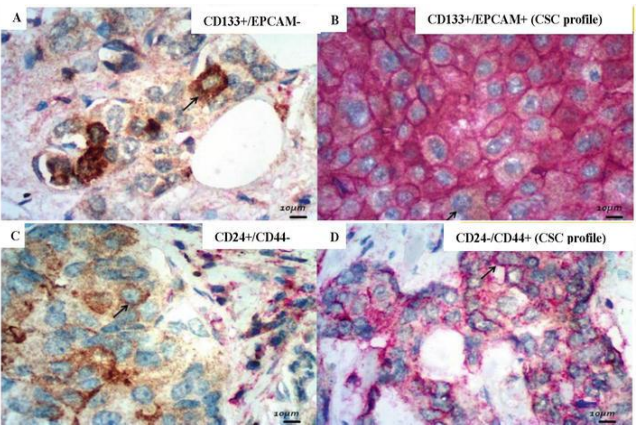
Figure 4.
Photomicrographies of double-marked plain stained presumptive CSC antibodies (400×, original praise, immunoperoxidase, and DAB). (A) CD133: cytoplasm positivity (immunoperoxidase); (B) EPCAM: sheath positiveness (DAB); (B) CD133+/EPCAM+: CSC profile (hopeless projectile: membrane alacrity to DAB and cytoplasm eagerness to immunoperoxidase at the same container); (C) CD24: cytoplasm alacrity (immunoperoxidase); (D) CD24−/CD44+: CSC profile (inky missile: membrane readiness only to DAB)
In a few studies, identification of a stem-container-like phenotype CD24 depressed/CD44 enriched has biased the innocent disease endurance, exceptionally in cases of early stages of feelings tumor, with more incidents of faraway metastasis and malignancy frequency after surgical and secondary situations. The presence of malignancy containers with eagerness for tumor stem-cell phenotype CD133/EPCAM has had a connection with poor overall endurance in bosom cancer, accompanying secondary therapeutic abandon [32]. For the moment, these presumptive stem-cell phenotypes appear expected independent prognostic determinants in feeling cancer. “Triple negative” feelings tumor and BRCA-1 mutant feelings tumor have existed associated with stem-container-like phenotype CD24 depressed/CD44 enriched. These presumptive stem-container markers can enhance possible future aims for new drugs from now on [30, 32].
5 Immunologic facets had a connection with Breast Cancer
In the circumstances of the tumor, the immune arrangement can restrain the lump growth apiece demolition of malignancy cells or hindrance of their consequence. On the other hand, the immune arrangement can imitate Cancer progression for one collection of swelling cells that are used to bear in an immunocompetent host or modify the lump atmosphere to facilitate the Cancer outgrowth [37]. Elevated levels of CD4+ supervisory T lymphocytes (Tregs) in the direction of many cancers are joined to poor prognosis. Tregs form an encouraging immunosuppressive microenvironment for the projection and progression of the cyst. On this habit, FOXP3 is articulated by the Tregs and may be discovered by immunohistochemistry. FOXP3 arranges induction and support of fortitude to self-antigens in normal containers, also this immunotolerance may be performed for one Tregs accompanying malignancy cell antigens [37, 38]. Another model of tumor container escape mechanism from the invulnerable arrangement is caspase-8 mutations present in “threefold negative” breast cancers and different stable diseased tumors. These mutations abolish the obliteration persuaded by cytotoxic lymphocytes CD8+ in tumoral containers [37, 39]. The activation of T lymphocytes by overseas antigens happens by contributing to major histocompatibility complex (MHC) irritant performance and co-verbalization of T-cell receptor (TCR). At the same time, kin of T-container transmembrane proteins CD28/B7, named “immune checkpoints”, produce co-inhibitory or co-stimulatory signals. The invulnerable checkpoints organize the T-cell immunotolerance to care for the tissues from unacceptable damage. Cancer containers may produce signals to restrict T-container operation, through cytotoxic T-lymphocyte associated irritant-4 (CTLA-4), prioritize container death-1 (PD-1), and allure ligands (PDL-1) [37, 40]. PD-1 is an inhibitory “invulnerable checkpoint” articulated on the surface of T-containers, B-containers, and NK-containers. When T-cells have mobilized by their TCR, the containers express at the same time PD-1, which is feasibility for the assaulted cell to escape from the invulnerable backlash (Figure 5). Cancer containers express the ligand PDL-1 on their surfaces, activating PD-1 of T-containers, emerging from the attack [37, 40].

Figure 5
Simplified diagrammatic drawing of PD-1/PDL-1 interactions in invulnerable reactions against cancer container. Tumoral antigens (Ag) are bestowed via T-container by the big histocompatibility complex (MHC) of dendritic containers. T-container perceives tumoral Ag via TCR (T-container receptor). Interaction Ag-TCR induces a helpful immune reaction against tumoral Ag. Though, skilled is a scape machine of malignancy cell from the T-container attack: interplay of set up death container ligands (PDL-1/2) articulated by cancer container accompanying PD-1 expressed by T-container prevent the T-cell operation. This view mechanism of malignancy container mimics the rule action to prevent invulnerable responses of T-containers against self-antigens. The law of immune analysis is the hindrance of PD-1/PDL-1 (extracted from [40]). PD-L1 verbalization has happened guide large lump proportion, high grade, extreme increase, estrogen receptor (ER)-negative status, and human epidermal development determinant receptor-2 (HER2)-positive rank in feelings malignancy. Survival in breast malignancy is with the order reversed related to PD-1/PDL-1 levels. PDL-1 verbalization increases cyst aggressiveness, exciting tumorigenesis, invasiveness, and skill to escape from cytotoxic T CD8+ lymphocyte attacks [39, 41]. The immunohistochemical evaluation of PDL-1 is proved in Figure 6.
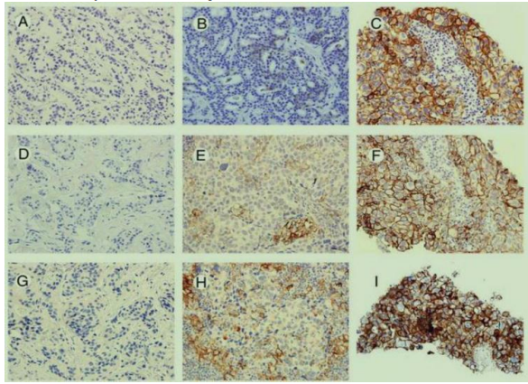
Figure 6
Examples of PDL-1 verbalization in conscience cancer utilizing 3 various antibodies: Dako 22C3 (D, E, and F), Ventana SP263 (G, H, and I), and BioCare RbM CAL10 (A, B, and C). PDL-1 cut is divided into 3 groups: nothing staining is negative, 1–49% of certain containers are considered “depressed PDL-1 verbalization” and 50% of more helpful cells are deliberate “extreme PDL-1 verbalization”. Examples of negative, low, and extreme PDL-1 verbalization are presented on A, B, and C for BioCare antibodies (culled from [42]).
Immune healings accompanying anti-CTLA-4 and antagonistic-PD1/antagonistic-PDL-1 powers have been hopeful for medicating various cancers. In breast malignancy, few researchers stated positive results in about 20% of bosom tumors in situations with these powers, for the most part, the “threefold negative” and HER-2 subtypes, for their higher antigenicity. In general, bosom tumors present lower immunogenicity than different cancers and breast malignancy containers commonly create an immunosuppressor carcinoma microenvironment by indicating [37, 43]. The demeanor of tumor permeating lymphocytes (TIL) in a few feeling cancers has had a connection with a friendly forecast, especially in “threefold negative” and HER-2 subtypes. TIL are made principally by T-cells CD3+/CD56 negative, that are either CD4+ or CD8+. A youth component of B-containers CD20+ and NK-cells concede possibility show. The attractiveness of TIL by cancer containers has had a connection with their expression of a few chemokines, like CXCL9 and CXCL13 [37, 44]. In “threefold negative” and HER-2 subtypes of conscience cancer, the demeanor of TIL is had connection with a better answer to neoadjuvant therapy, also neoadjuvant situation grant permission modify the swelling microenvironment to engage TIL to carcinoma site. Furthermore, when the TIL are not brought a suggestion of correction neoadjuvance, it is exhibitive for bad forecast [44].
6. Advanced stage bosom malignancy: considerations undercurrent approach and future outlooks
Metastatic feelings cancer is deliberately unchangeable at this time with no remedies. Therapy of metastatic affliction aims to guarantee quality of existence, relief of symptoms, and extension of the patient continuation. Advanced stage affliction is becoming more and more never-ending, conditional sequential therapies, accompanying made-to-order approach than the inception of breast malignancy [8]. Systemic analysis is repeatedly the first choice of metastatic affliction. Before the new healing resolution, it is necessary to believe the former situations. If possible, it is recommended to reconsider the histologic facial characteristics and microscopic subtype status of the metastatic wound through a new medical checkup, accompanying a new immunohistochemical study for hormonal receptor and HER-2 status. Some studies stated just before 40% of discrepancies of metastatic lesion histologic appearance and microscopic subtype rank versus basic cyst histologic and immunohistochemical facets [45]. The metastatic disease healing selections follow positive aims to hit the neoplastic containers. Thereby, the expression of hormonal receptors for one metastatic wound is important for endocrine therapy. Endocrine drugs involve tamoxifen, aromatase inhibitors, fulvestrant and progestins. The use of these drugs in often major accompanying hormone receptor definite rank has illustrated an increase of free of ailment continuation in various studies [8, 45]. Furthermore, generation after baby boom of drugs that restrict the cyclin-dependant kinase (CDK) has been profitable in the extension of innocent ailment survival in luminal subtype HER-2 negative metastatic ailment. CDK4/6 is a holoenzyme being the reason for various extracellular indicating pathways to cell era changes. CDK4/6 fosforilates and inactivates retinoblastoma cyst suppressor protein (Rb). Extracellular signals organize the expression of cyclins and CDK inhibitors, like p16INK4a [46]. In human malignancy, this track is dysregulated by either the overexpression of cyclin D1, the misfortune of p16Ink4a, the metamorphosis of CDK4 to an Ink4-refractory state, or the misfortune of Rb itself. The basic goal of CDK4 is the Rb protein, though this holoenzyme either can phosphorylate determinants involved in container distinction moving their transcriptional endeavor, apoptotic factors moving their project, and additional determinants that can directly influence mitochondrial function [8, 46, 47]. Therefore, CDK inhibitors act in carcinoma microenvironment, obstructing Rb phosphorilation and superior to cell era exit. Moreover, CDK have kinase venture towards SPOP, an ubiquitin protein that communicates accompanying PDL-1. CDK inhibitors lead to restriction of SPOP phosphorilation accompanying barrier of PDL-1 and provocation to PD-1 expression by T-containers, appealing to T-container combination to the tumor. In this way, the linked use of CDK inhibitors and PDL-1/PD-1 inhibitors can be hopeful, needing more future studies [46, 47, 48]. For the moment, hormonal receptors and HER-2 rank are the few substantiated microscopic marks of dispassionate importance on metastatic conscience malignancy nearing through a destructive agent and endocrine therapy. For HER-2 certain metastatic afflictions, antagonistic-HER-2 situation with trastuzumab is traditional and is urged as shortly as likely. Immune therapy is not patterned for metastatic conscience malignancy, because metastatic breast ailment is well various. Though it is a hopeful therapy for the future, it also aims for microscopic analyses, which enhance more direct accompanying findings of novel pathways and mutations by new studies to be grown [8]. A take up of main biomarkers of clinicopathologic significance for feelings cancer administration is proved in Table 2 and a suggestion of a treasure for clinicopathologic evaluation of bosom malignancy is bestowed in Table 3.
| Biomarker | Detection Technique | Nature | Clinicopathologic Importance |
|---|---|---|---|
| Hormonal Receptors / HER2 | IHC / FISH | Biomarkers of molecular subtypes of breast cancer | Targets for endocrine and anti-HER2 therapies; prognostic predictors |
| BRCA1 / BRCA2 | PCR sequencing | Biomarker of hereditary breast cancer | Target for PARP inhibitors; indication for screening of other malignancies |
| CD24, CD44, CD133, EPCAM | IHC | Putative stem-cell biomarkers | Prediction of poor prognosis, tumor progression risk, and survival reduction |
| PD-1 / PDL-1 | IHC | Biomarker of possible inhibited immune response of T-cells against cancer cells | Target for immunotherapy using PD-1/PDL-1 antagonists |
| TILs (Tumor-Infiltrating Lymphocytes) | Histologic assessment and IHC | Marker of better cellular-mediated immune response against cancer cells | Predicts better therapeutic response, especially to neoadjuvant therapies |
1HC = Immunohistochemistry.
2TILs = Tumoral permeating lymphocytes.
Table 2: Summary of Main Biomarkers of Clinicopathologic Importance for Breast Cancer Management
| Step | Evaluation Criteria | Tests/Methods | Clinical Significance |
|---|---|---|---|
| 1. Patient History & Physical Exam | Family history, risk factors, symptoms (lump, pain, nipple discharge, etc.) | Clinical breast exam (CBE) | Identifies high-risk patients and symptomatic cases |
| 2. Imaging Studies | Suspicious findings in clinical exam | Mammography, ultrasound, MRI | Differentiates benign vs. malignant lesions, detects non-palpable tumors |
| 3. Biopsy & Pathologic Diagnosis | Suspicious imaging findings | Core needle biopsy, fine-needle aspiration, excisional biopsy | Confirms malignancy and histologic subtype |
| 4. Histopathologic Evaluation | Tumor type, grade, lymphovascular invasion | H&E staining, Nottingham grading system | Determines tumor aggressiveness and prognosis |
| 5. Immunohistochemistry (IHC) & Biomarker Analysis | Hormone receptor (ER, PR), HER2 status, Ki-67 proliferation index | IHC, FISH (for HER2) | Guides targeted therapy (hormonal, HER2-directed) |
| 6. Molecular Testing (if indicated) | High-risk or triple-negative cases | Oncotype DX, Mammaprint, BRCA mutation testing | Predicts recurrence risk, eligibility for chemotherapy or genetic counseling |
| 7. Staging & Metastatic Workup | Tumor size, nodal involvement, distant metastases | TNM classification, CT, PET-CT, bone scan | Determines stage and treatment plan |
| 8. Multidisciplinary Tumor Board Review | Integrative case discussion | Oncology, surgery, radiology, pathology review | Optimizes treatment strategy based on case-specific factors |
| 9. Treatment Planning | Surgery, radiation, systemic therapy | Breast-conserving surgery (BCS), mastectomy, neoadjuvant/adjuvant therapy | Individualized treatment approach for best outcomes |
| 10. Follow-up & Surveillance | Recurrence risk, long-term monitoring | Regular clinical exams, imaging, tumor markers | Ensures early detection of recurrence or new malignancies |
Table 3: Proposal of an Algorithm for Clinicopathologic Evaluation of Breast Cancer.
This study engages an assortedmethods approach, joining ancient analysis accompanying modern research judgment to explore the progress of the study of plants from classical methods to up-to-date novelties. A comprehensive essay review was transported using peer-inspected journals, books, and dispassionate studies connecting the past centennial. Additionally, concerning qualities not quantities interviews with study of plants masters and determinable data from current mechanics advancements, to a degree mathematical study of plants and artificial intelligence uses, were resolved. Comparative analysis was used to evaluate the impact of creative techniques on demonstrative veracity and patient outcomes.
The judgments display a meaningful transition from established histopathology patterns to advanced microscopic and mathematical study of plant techniques. Key results contain: Improved Diagnostic Accuracy: Digital image and AI-assisted study of plants have raised the accuracy of disease disease by 30-40% distinguished to classical plans. Faster Turnaround Time: The unification of mechanization and computational pathology has weakened demonstrative delays by approximately 50%. Enhanced Disease Characterization: Molecular study of plants and biomarker study have simplified early disease discovery, specifically in oncology.
Adoption Challenges: Despite these advancements, monetary and infrastructural restraints deter widespread exercise, specifically in resource-restricted backgrounds.
The conversion of pathology over age has happened driven by concerning details progress that embellish diagnostic competencies. The change from chaste techniques to a degree of hematoxylin and eosin (H&E) staining and manual microscopy, to new digital and microscopic study of plants has transformed disease discovery and situation planning. AI-located study of plants has imported unprecedented veracity and effectiveness, reducing human wrongs and permissive big data study. However, challenges in the way that the high cost of electronics, the need for specific preparation, and ethical concerns concerning AI-compelled diagnoses must be addressed to guarantee impartial ratification. Future research should devote effort to something mixing these innovations seamlessly into routine dispassionate practice and expanding economical solutions for depressed-means settings.
Pathology has sustained an extraordinary transformation, transitioning from normal histopathological methods to leading microscopic and computational approaches. The integration of AI, the mathematical study of plants, and microscopic diagnostics has considerably embellished demonstrative accuracy and efficiency. While this progress holds excellent promise, challenges had connection with cost, training, and righteous concerns must be sent to guarantee widespread maintenance. Continued research and mechanics cultivation will be key to maintaining the study of plants at the prominence of healing change, ultimately reconstructing patient care and clinical consequences.
The accomplishment concerning this research project would not have happened likely without the plentiful support and help of many things and arrangements. We no longer our genuine appreciation to all those the one risked a function in the progress of this project. I herewith acknowledge that: I have no economic or added individual interests, straightforwardly or obliquely, in some matter that conceivably influence or bias my trustworthiness as a journalist concerning this Manuscript
The authors declare that they have no conflicts of interest.
No external funding for a project was taken to assist with the preparation of this manuscript
“I would like to express my sincere gratitude to Ms. Sarah Jacobs, Editorial Coordinator of the IJCCR, for her excellent collaboration and exceptional support throughout the entire process, from article submission and peer review to financial matters and successful publication. Everything went very quickly and smoothly. I would also like to thank the reviewers, whose comments were helpful and contributed to an even better article. I wholeheartedly recommend this deservedly renowned journal to my colleagues for publication.” Best regards, Andreas Schapowal.

Dear Editorial Team, Clinical Medical Reviews and Reports. My experience with the journal was highly positive. The peer-review process was rigorous, constructive, and completed in a timely manner. The reviewers provided valuable comments that helped improve the quality and clarity of our manuscript. The editorial office was professional, responsive, and supportive throughout all stages of the publication process. Communication was clear and efficient, and any questions were addressed promptly. Overall, I found the journal to maintain high scientific standards and an excellent publication workflow. I would be pleased to consider submitting future work to this journal. Best wishes from, Elena Popa.

It was my pleasure to submit my testimonial concerning the Reviewer Board of our Scientific Journal “Brain and Neurological Disorders”. The Reviewers focused on some modifications and their contribution was helpful. The ladies of our Editorial Office were also supported my efforts. It was my honor to have such a co-operation and I am looking forward for more collaboration.

Dear Grace Pierce, Editorial Coordinator of Journal of Clinical Research and Reports, Thank you for the speedy and efficient peer review process. I appreciate the fact that your peer reviewers do not take months to respond like with some other journals. I would also like to thank the editorial office for responding quickly to my questions. It is an excellent journal. I plan to submit more manuscripts in the future. Best wishes from, Robert W. McGee

Dear Grace Pierce, Editorial Coordinator of Journal of Clinical Research and Reports, Working with you and your team on our recent publication in JCRR has been a truly wonderful and enjoyable experience. The responses were prompt, and the reviewers were patient, constructive, and highly professional. One reviewer in particular gave me the feeling that a professor was carefully reading and commenting on my coursework, which was deeply touching. The entire process was straightforward and hassle‑free, with no tedious online forms to complete. I highly recommend this journal. Best wishes from, DR Aibing Rao, Head of R&D

I Appreciate the Opportunity to Share my Experience with the Journal of Clinical Research and Reports. The peer review process was timely and constructive, and the feedback provided helped improve the quality of our manuscript. The editorial office was professional, responsive, and supportive throughout the process, ensuring smooth communication and efficient handling of the submission. Overall, it was a positive experience collaborating with your team.

Dear Mercy Grace, Editorial Coordinator of Obstetrics Gynecology and Reproductive Sciences, We would like to express our gratitude for your help at all stages of publishing and editing the article. The editors of the magazine answer all the necessary questions and help at every stage. We will definitely continue to cooperate and publish other works in the Obstetrics Gynecology and Reproductive Sciences! Best wishes from, Alla Konstantinovna Politova,

Dear Maria Emerson, Editorial Coordinator of International Journal of Clinical Case Reports and Reviews, What distinguishes International Journal of Clinical Case Report and Review is not only the scientific rigor of its publications, but the intellectual climate in which research is evaluated. The submission process is refreshingly free of unnecessary formal barriers and bureaucratic rituals that often complicate academic publishing without adding real value. The peer-review system is demanding yet constructive, guided by genuine scientific dialogue rather than hierarchical or authoritarian attitudes. Reviewers act as collaborators in improving the manuscript, not as gatekeepers imposing arbitrary standards. This journal offers a rare balance: high methodological standards combined with a respectful, transparent, and supportive editorial approach. In an era where publishing can feel more burdensome than research itself, this platform restores the original purpose of peer review — to refine ideas, not to obstruct them Prof. Perlat Kapisyzi, FCCP PULMONOLOGIST AND THORACIC IMAGING.

Dear Reader: We have published several articles in the Auctores Publishing, LLC, journal, Clinical Medical Reviews and Reports in recent years (CMRR). This is an ‘open access’ journal and the following are our observations. From the initial invitation to submit an article, to the final edits of galley proofs, we have found CMRR personnel to be professional, responsive, rapid and thorough. This entire process begins with Catherine Mitchell, Editorial Coordinator. She is simply outstanding, and, I believe, unparalleled in her capacity. I cannot imagine a more responsive and dedicated Editorial Coordinator. As I read the dates and timing of her correspondence with us, it seems that she never sleeps. I hope Auctores Publishing, LLC, appreciates her efforts as much as these authors do. Thank you to Auctores Publishing, LLC, to the Editorial Staff/Board, and to Catherine Mitchell from a grateful author(s).

Dear Maria Emerson, Editorial Coordinator of - Journal of Clinical Research and Reports. ''I am pleased to provide this testimonial following the publication of our recent case report in this journal. The peer review process was rigorous, constructive, thorough, and conducted in a timely manner. The reviewers’ comments were thoughtful, detailed, and highly constructive, contributing substantially to the refinement, clarity, and scientific robustness of our manuscript. The process was conducted with professionalism and academic integrity throughout. The support provided by the editorial office was exemplary. Communication was consistently prompt, clear, and courteous at all stages of the submission and publication process. The editorial team demonstrated a high level of organization and responsiveness, ensuring that all queries were addressed efficiently and that the process remained transparent and well-coordinated. The overall quality of the journal is reflected in its strong editorial standards, commitment to scientific excellence, and dedication to publishing clinically meaningful research. It has been a privilege to publish our work in this journal, and we would welcome the opportunity to contribute further in the future.'' Best wishes from, Dr. Efstratios Trogkanis, Cardiologist.

Dear Grace Pierce, Editorial Coordinator of the journal IJCCR, I had a very positive experience with Auctores - Journal throughout the publication process. The Editorial Team was highly responsive, professional, and supportive at every stage. I would like to extend my sincere thanks to the Editor: Grace Pierce, for her guidance and assistance. The peer-review process was smooth and constructive, helping improve the quality of my work. I would gladly recommend Auctores Journal to fellow researchers and authors. Dr. SABITA SINHA, Medical Oncologist, MD (Electro Homeopathy).

Dear Mayra Duenas, Editorial Coordinator of the journal IJCCR, I write here a little on my experience as an author submitting to the International Journal of Clinical Case Reports and Reviews (IJCCR). This was my first submission to IJCCR and my manuscript was inherently an outsider’s effort. It attempted to broadly identify and then make some sense of life’s under-appreciated mysteries. I initially had responded to a request for possible submissions. I then contacted IJCCR with a tentative topic for a manuscript. They quickly got back with an approval for the submission, but with a particular requirement that it be medically relevant. I then put together a manuscript and submitted it. After the usual back-and-forth over forms and formality, the manuscript was sent off for reviews. Within 2 weeks I got back 4 reviews which were both helpful and also surprising. Surprising in that the topic was somewhat foreign to medical literature. My subsequent updates in response to the reviewer comments went smoothly and in short order I had a series of proofs to evaluate. All in all, the whole publication process seemed outstanding. It was both helpful in terms of the paper’s content and also in terms of its efficient and friendly communications. Thank you all very much. Sincerely, Ted Christopher, Rochester, NY.

International Journal of Clinical Case Reports and Reviews is a high quality journal that has a clear and concise submission process. The peer review process was comprehensive and constructive. Support from the editorial office was excellent, since the administrative staff were responsive. The journal provides a fast and timely publication timeline.

Dear Cecilia Lilly, Editorial Coordinator, Endocrinology and Disorders, Thank you so much for your quick response regarding reviewing and all process till publishing our manuscript entitled: Prevalence of Pre-Diabetes and its Associated Risk Factors Among Nile College Students, Sudan. Best regards, Dr Mamoun Magzoub.

Dear Editorial Team, Clinical Cardiology and Cardiovascular Interventions. I am really grateful for the peers review; their feedback gave me the opportunity to reflect on the message and impact of my work and to ameliorate the article. The editors did a great job in addition by encouraging me to continue with the process of publishing.

Dear Mayra Duenas, Editorial Coordinator of ‘International Journal of Clinical Case Reports and Reviews Herewith I confirm an optimal peer review process and a great support of the editorial office of the present journal

Dear Maria Emerson, Editorial Coordinator of ‘International Journal of Clinical Case Reports and Reviews’, I appreciate the opportunity to publish my article with your journal. The editorial office provided clear communication during the submission and review process, and I found the overall experience professional and constructive. Best regards, Elena Salvatore.

Dear Editorial Team, Journal-Clinical Cardiology and Cardiovascular Interventions, “Publishing my article with Clinical Cardiology and Cardiovascular Interventions has been a highly positive experience. The peer-review process was rigorous yet supportive, offering valuable feedback that strengthened my work. The editorial team demonstrated exceptional professionalism, prompt communication, and a genuine commitment to maintaining the highest scientific standards. I am very pleased with the publication quality and proud to be associated with such a reputable journal.” Warm regards, Dr. Mahmoud Kamal Moustafa Ahmed

Dear Editorial Team, Clinical Cardiology and Cardiovascular Interventions. It was truly a rewarding experience to work with the journal “Clinical Cardiology and Cardiovascular Interventions”. The peer review process was insightful and encouraging, helping us refine our work to a higher standard. The editorial office offered exceptional support with prompt and thoughtful communication. I highly value the journal’s role in promoting scientific advancement and am honored to be part of it. Best regards, Meng-Jou Lee, MD, Department of Anesthesiology, National Taiwan University Hospital.

Dear Maria Emerson, Editorial Coordinator, Journal of Clinical Research and Reports. Thank you for publishing our case report: "Clinical Case of Effective Fetal Stem Cells Treatment in a Patient with Autism Spectrum Disorder" within the "Journal of Clinical Research and Reports" being submitted by the team of EmCell doctors from Kyiv, Ukraine. We much appreciate a professional and transparent peer-review process from Auctores. All research Doctors are so grateful to your Editorial Office and Auctores Publishing support! I amiably wish our article publication maintained a top quality of your International Scientific Journal. My best wishes for a prosperity of the Journal of Clinical Research and Reports. Hope our scientific relationship and cooperation will remain long lasting. Thank you very much indeed. Kind regards, Dr. Andriy Sinelnyk Cell Therapy Center EmCell

I recommend without hesitation submitting relevant papers on medical decision making to the International Journal of Clinical Case Reports and Reviews. I am very grateful to the editorial staff. Maria Emerson was a pleasure to communicate with. The time from submission to publication was an extremely short 3 weeks. The editorial staff submitted the paper to three reviewers. Two of the reviewers commented positively on the value of publishing the paper. The editorial staff quickly recognized the third reviewer’s comments as an unjust attempt to reject the paper. I revised the paper as recommended by the first two reviewers.

Dear Chrystine Mejia, Editorial Coordinator, Journal of Neurodegeneration and Neurorehabilitation. “The peer review process was efficient and constructive, and the editorial office provided excellent communication and support throughout. The journal ensures scientific rigor and high editorial standards, while also offering a smooth and timely publication process. We sincerely appreciate the work of the editorial team in facilitating the dissemination of innovative approaches such as the Bonori Method.” Best regards, Dr. Matteo Bonori.

Dear Maria Emerson, Editorial Coordinator, we have deeply appreciated the professionalism demonstrated by the International Journal of Clinical Case Reports and Reviews. The reviewers have extensive knowledge of our field and have been very efficient and fast in supporting the process. I am really looking forward to further collaboration. Thanks. Best regards, Dr. Claudio Ligresti

Dear Ms. Mayra Duenas, Editorial Coordinator, International Journal of Clinical Case Reports and Reviews. “The International Journal of Clinical Case Reports and Reviews represented the “ideal house” to share with the research community a first experience with the use of the Simeox device for speech rehabilitation. High scientific reputation and attractive website communication were first determinants for the selection of this Journal, and the following submission process exceeded expectations: fast but highly professional peer review, great support by the editorial office, elegant graphic layout. Exactly what a dynamic research team - also composed by allied professionals - needs!" From, Chiara Beccaluva, PT - Italy.

Dear Clarissa Eric, Editorial Coordinator, Journal of Clinical Case Reports and Studies, Auctores Publishing LLC, USA Office: +1-(302)-520-2644. I would like to express my sincere appreciation for the efficient and professional handling of my case report by the ‘Journal of Clinical Case Reports and Studies’. The peer review process was not only fast but also highly constructive—the reviewers’ comments were clear, relevant, and greatly helped me improve the quality and clarity of my manuscript. I also received excellent support from the editorial office throughout the process. Communication was smooth and timely, and I felt well guided at every stage, from submission to publication. The overall quality and rigor of the journal are truly commendable. I am pleased to have published my work with Journal of Clinical Case Reports and Studies, and I look forward to future opportunities for collaboration. Sincerely, Aline Tollet, UCLouvain.

Dear Chrystine Mejia, Editorial Coordinator, Journal of Neurodegeneration and Neurorehabilitation, Auctores Publishing LLC, We would like to thank the editorial team for the smooth and high-quality communication leading up to the publication of our article in the Journal of Neurodegeneration and Neurorehabilitation. The reviewers have extensive knowledge in the field, and their relevant questions helped to add value to our publication. Kind regards, Dr. Ravi Shrivastava.

Dear Clarissa Eric, Editorial Coordinator, Journal of Clinical Case Reports and Studies, I would like to express my deep admiration for the exceptional professionalism demonstrated by your journal. I am thoroughly impressed by the speed of the editorial process, the substantive and insightful reviews, and the meticulous preparation of the manuscript for publication. Additionally, I greatly appreciate the courteous and immediate responses from your editorial office to all my inquiries. Best Regards, Dariusz Ziora

Dear Ashley Rosa, Editorial Coordinator, International Journal of Clinical Case Reports and Reviews, Auctores Publishing LLC. Thank you for publishing our article, Exploring Clozapine's Efficacy in Managing Aggression: A Multiple Single-Case Study in Forensic Psychiatry in the international journal of clinical case reports and reviews. We found the peer review process very professional and efficient. The comments were constructive, and the whole process was efficient. On behalf of the co-authors, I would like to thank you for publishing this article. With regards, Dr. Jelle R. Lettinga.

Dear Jessica Magne, Editorial Coordinator, Clinical Cardiology and Cardiovascular Interventions, Auctores Publishing LLC. The peer review process of the journal of Clinical Cardiology and Cardiovascular Interventions was excellent and fast, as was the support of the editorial office and the quality of the journal. Kind regards Walter F. Riesen Prof. Dr. Dr. h.c. Walter F. Riesen.

Dear Erin Aust, Editorial Coordinator, Journal of General Medicine and Clinical Practice. We are pleased to share our experience with the “Journal of General Medicine and Clinical Practice”, following the successful publication of our article. The peer review process was thorough and constructive, helping to improve the clarity and quality of the manuscript. We are especially thankful to Ms. Erin Aust, the Editorial Coordinator, for her prompt communication and continuous support throughout the process. Her professionalism ensured a smooth and efficient publication experience. The journal upholds high editorial standards, and we highly recommend it to fellow researchers seeking a credible platform for their work. Best wishes By, Dr. Rakhi Mishra.

Dr Hala Al Shaikh This is to acknowledge that the peer review process for the article ’ A Novel Gnrh1 Gene Mutation in Four Omani Male Siblings, Presentation and Management ’ sent to the International Journal of Clinical Case Reports and Reviews was quick and smooth. The editorial office was prompt with easy communication.

Dear Agrippa Hilda, Editorial Coordinator, Journal of Neuroscience and Neurological Surgery. The entire process including article submission, review, revision, and publication was extremely easy. The journal editor was prompt and helpful, and the reviewers contributed to the quality of the paper. Thank you so much! Eric Nussbaum, MD

Dear Ashley Rosa, Editorial Coordinator, International Journal of Clinical Case Reports and Reviews. Many thanks for publishing this manuscript after I lost confidence the editors were most helpful, more than other journals Best wishes from, Susan Anne Smith, PhD. Australian Breastfeeding Association.

Dear Maria Emerson, Editorial Coordinator, International Journal of Clinical Case Reports and Reviews, Auctores Publishing LLC. I am delighted to have published our manuscript, "Acute Colonic Pseudo-Obstruction (ACPO): A rare but serious complication following caesarean section." I want to thank the editorial team, especially Maria Emerson, for their prompt review of the manuscript, quick responses to queries, and overall support. Yours sincerely Dr. Victor Olagundoye.

Dear Jessica Magne, Editorial Coordinator, Clinical Cardiology and Cardiovascular Interventions, Auctores Publishing LLC. I appreciate the journal (JCCI) editorial office support, the entire team leads were always ready to help, not only on technical front but also on thorough process. Also, I should thank dear reviewers’ attention to detail and creative approach to teach me and bring new insights by their comments. Surely, more discussions and introduction of other hemodynamic devices would provide better prevention and management of shock states. Your efforts and dedication in presenting educational materials in this journal are commendable. Best wishes from, Farahnaz Fallahian.

Dear Ashley Rosa, Editorial Coordinator of the journal - Psychology and Mental Health Care. " The process of obtaining publication of my article in the Psychology and Mental Health Journal was positive in all areas. The peer review process resulted in a number of valuable comments, the editorial process was collaborative and timely, and the quality of this journal has been quickly noticed, resulting in alternative journals contacting me to publish with them." Warm regards, Susan Anne Smith, PhD. Australian Breastfeeding Association.

Dear Jessica, and the super professional team of the ‘Clinical Cardiology and Cardiovascular Interventions’ I am sincerely grateful to the coordinated work of the journal team for the no problem with the submission of my manuscript: “Cardiometabolic Disorders in A Pregnant Woman with Severe Preeclampsia on the Background of Morbid Obesity (Case Report).” The review process by 5 experts was fast, and the comments were professional, which made it more specific and academic, and the process of publication and presentation of the article was excellent. I recommend that my colleagues publish articles in this journal, and I am interested in further scientific cooperation. Sincerely and best wishes, Dr. Oleg Golyanovskiy.

To Dear Erin Aust – Editorial Coordinator of Journal of General Medicine and Clinical Practice! I declare that I am absolutely satisfied with your work carried out with great competence in following the manuscript during the various stages from its receipt, during the revision process to the final acceptance for publication. Thank Prof. Elvira Farina

My article, titled 'No Way Out of the Smartphone Epidemic Without Considering the Insights of Brain Research,' has been republished in the International Journal of Clinical Case Reports and Reviews. The review process was seamless and professional, with the editors being both friendly and supportive. I am deeply grateful for their efforts.

We found the peer review process quick and positive in its input. The support from the editorial officer has been very agile, always with the intention of improving the article and taking into account our subsequent corrections.

Dear Jessica Magne, with gratitude for the joint work. Fast process of receiving and processing the submitted scientific materials in “Clinical Cardiology and Cardiovascular Interventions”. High level of competence of the editors with clear and correct recommendations and ideas for enriching the article.

I thank the ‘Journal of Clinical Research and Reports’ for accepting this article for publication. This is a rigorously peer reviewed journal which is on all major global scientific data bases. I note the review process was prompt, thorough and professionally critical. It gave us an insight into a number of important scientific/statistical issues. The review prompted us to review the relevant literature again and look at the limitations of the study. The peer reviewers were open, clear in the instructions and the editorial team was very prompt in their communication. This journal certainly publishes quality research articles. I would recommend the journal for any future publications.

"I am grateful for the opportunity of contributing to [International Journal of Clinical Case Reports and Reviews] and for the rigorous review process that enhances the quality of research published in your esteemed journal. I sincerely appreciate the time and effort of your team who have dedicatedly helped me in improvising changes and modifying my manuscript. The insightful comments and constructive feedback provided have been invaluable in refining and strengthening my work".

To Dear Erin Aust, I would like to express my heartfelt appreciation for the opportunity to have my work published in this esteemed journal. The entire publication process was smooth and well-organized, and I am extremely satisfied with the final result. The Editorial Team demonstrated the utmost professionalism, providing prompt and insightful feedback throughout the review process. Their clear communication and constructive suggestions were invaluable in enhancing my manuscript, and their meticulous attention to detail and dedication to quality are truly commendable. Additionally, the support from the Editorial Office was exceptional. From the initial submission to the final publication, I was guided through every step of the process with great care and professionalism. The team's responsiveness and assistance made the entire experience both easy and stress-free. I am also deeply impressed by the quality and reputation of the journal. It is an honor to have my research featured in such a respected publication, and I am confident that it will make a meaningful contribution to the field.

I would like to express my sincere gratitude for the support and efficiency provided by the editorial office throughout the publication process of my article, “Delayed Vulvar Metastases from Rectal Carcinoma: A Case Report.” I greatly appreciate the assistance and guidance I received from your team, which made the entire process smooth and efficient. The peer review process was thorough and constructive, contributing to the overall quality of the final article. I am very grateful for the high level of professionalism and commitment shown by the editorial staff, and I look forward to maintaining a long-term collaboration with the International Journal of Clinical Case Reports and Reviews.

Dear Agrippa Hilda- Editorial Coordinator of Journal of Neuroscience and Neurological Surgery, "The peer review process was very quick and of high quality, which can also be seen in the articles in the journal. The collaboration with the editorial office was very good."

I would like to offer my testimony in the support. I have received through the peer review process and support the editorial office where they are to support young authors like me, encourage them to publish their work in your esteemed journals, and globalize and share knowledge globally. I really appreciate your journal, peer review, and editorial office.

My experience publishing in International Journal of Clinical Case Reports and Reviews was exceptional. I Come forth to Provide a Testimonial Covering the Peer Review Process and the editorial office for the Professional and Impartial Evaluation of the Manuscript.

My experience publishing in Psychology and Mental Health Care was exceptional. The peer review process was rigorous and constructive, with reviewers providing valuable insights that helped enhance the quality of our work. The editorial team was highly supportive and responsive, making the submission process smooth and efficient. The journal's commitment to high standards and academic rigor makes it a respected platform for quality research. I am grateful for the opportunity to publish in such a reputable journal.

My Testimonial Covering as fellowing: Lin-Show Chin. The peer reviewers process is quick and effective, the supports from editorial office is excellent, the quality of journal is high. I would like to collabroate with Internatioanl journal of Clinical Case Reports and Reviews.

Dealing with The Journal of Neurology and Neurological Surgery was very smooth and comprehensive. The office staff took time to address my needs and the response from editors and the office was prompt and fair. I certainly hope to publish with this journal again.Their professionalism is apparent and more than satisfactory. Susan Weiner

Dear Editorial Coordinator of the Journal of Nutrition and Food Processing! "I would like to thank the Journal of Nutrition and Food Processing for including and publishing my article. The peer review process was very quick, movement and precise. The Editorial Board has done an extremely conscientious job with much help, valuable comments and advices. I find the journal very valuable from a professional point of view, thank you very much for allowing me to be part of it and I would like to participate in the future!”

Dear Monica Gissare, - Editorial Coordinator of Nutrition and Food Processing. ¨My testimony with you is truly professional, with a positive response regarding the follow-up of the article and its review, you took into account my qualities and the importance of the topic¨.

Dear Dr. Jessica Magne, Editorial Coordinator 0f Clinical Cardiology and Cardiovascular Interventions, I hope this message finds you well. I want to express my utmost gratitude for your excellent work and for the dedication and speed in the publication process of my article titled "Navigating Innovation: Qualitative Insights on Using Technology for Health Education in Acute Coronary Syndrome Patients." I am very satisfied with the peer review process, the support from the editorial office, and the quality of the journal. I hope we can maintain our scientific relationship in the long term.

Clinical Cardiology and Cardiovascular Interventions, I would like to express my sincerest gratitude for the trust placed in our team for the publication in your journal. It has been a true pleasure to collaborate with you on this project. I am pleased to inform you that both the peer review process and the attention from the editorial coordination have been excellent. Your team has worked with dedication and professionalism to ensure that your publication meets the highest standards of quality. We are confident that this collaboration will result in mutual success, and we are eager to see the fruits of this shared effort.

The peer reviewers process is quick and effective, the supports from editorial office is excellent, the quality of journal is high. I would like to collabroate with Internatioanl journal of Clinical Case Reports and Reviews journal clinically in the future time.

Clinical Cardiology and Cardiovascular Interventions, we deeply appreciate the interest shown in our work and its publication. It has been a true pleasure to collaborate with you. The peer review process, as well as the support provided by the editorial office, have been exceptional, and the quality of the journal is very high, which was a determining factor in our decision to publish with you.

Clinical Cardiology and Cardiovascular Interventions I testity the covering of the peer review process, support from the editorial office, and quality of the journal.

Dear editorial department: On behalf of our team, I hereby certify the reliability and superiority of the International Journal of Clinical Case Reports and Reviews in the peer review process, editorial support, and journal quality. Firstly, the peer review process of the International Journal of Clinical Case Reports and Reviews is rigorous, fair, transparent, fast, and of high quality. The editorial department invites experts from relevant fields as anonymous reviewers to review all submitted manuscripts. These experts have rich academic backgrounds and experience, and can accurately evaluate the academic quality, originality, and suitability of manuscripts. The editorial department is committed to ensuring the rigor of the peer review process, while also making every effort to ensure a fast review cycle to meet the needs of authors and the academic community. Secondly, the editorial team of the International Journal of Clinical Case Reports and Reviews is composed of a group of senior scholars and professionals with rich experience and professional knowledge in related fields. The editorial department is committed to assisting authors in improving their manuscripts, ensuring their academic accuracy, clarity, and completeness. Editors actively collaborate with authors, providing useful suggestions and feedback to promote the improvement and development of the manuscript. We believe that the support of the editorial department is one of the key factors in ensuring the quality of the journal. Finally, the International Journal of Clinical Case Reports and Reviews is renowned for its high- quality articles and strict academic standards. The editorial department is committed to publishing innovative and academically valuable research results to promote the development and progress of related fields. The International Journal of Clinical Case Reports and Reviews is reasonably priced and ensures excellent service and quality ratio, allowing authors to obtain high-level academic publishing opportunities in an affordable manner. I hereby solemnly declare that the International Journal of Clinical Case Reports and Reviews has a high level of credibility and superiority in terms of peer review process, editorial support, reasonable fees, and journal quality. Sincerely, Rui Tao.

“The peer review process of JPMHC is quick and effective. Authors are benefited by good and professional reviewers with huge experience in the field of psychology and mental health. The support from the editorial office is very professional. People to contact to are friendly and happy to help and assist any query authors might have. Quality of the Journal is scientific and publishes ground-breaking research on mental health that is useful for other professionals in the field”.

Dr.Tania Muñoz, My experience as researcher and author of a review article in The Journal Clinical Cardiology and Interventions has been very enriching and stimulating. The editorial team is excellent, performs its work with absolute responsibility and delivery. They are proactive, dynamic and receptive to all proposals. Supporting at all times the vast universe of authors who choose them as an option for publication. The team of review specialists, members of the editorial board, are brilliant professionals, with remarkable performance in medical research and scientific methodology. Together they form a frontline team that consolidates the JCCI as a magnificent option for the publication and review of high-level medical articles and broad collective interest. I am honored to be able to share my review article and open to receive all your comments.

I am delighted to publish our manuscript entitled "A Perspective on Cocaine Induced Stroke - Its Mechanisms and Management" in the Journal of Neuroscience and Neurological Surgery. The peer review process, support from the editorial office, and quality of the journal are excellent. The manuscripts published are of high quality and of excellent scientific value. I recommend this journal very much to colleagues.

I would like to give my testimony in the support I have got by the peer review process and to support the editorial office where they were of asset to support young author like me to be encouraged to publish their work in your respected journal and globalize and share knowledge across the globe. I really give my great gratitude to your journal and the peer review including the editorial office.

I am very pleased to serve as EBM of the journal, I hope many years of my experience in stem cells can help the journal from one way or another. As we know, stem cells hold great potential for regenerative medicine, which are mostly used to promote the repair response of diseased, dysfunctional or injured tissue using stem cells or their derivatives. I think Stem Cell Research and Therapeutics International is a great platform to publish and share the understanding towards the biology and translational or clinical application of stem cells.

We are grateful for this opportunity to provide a glowing recommendation to the Journal of Psychiatry and Psychotherapy. We found that the editorial team were very supportive, helpful, kept us abreast of timelines and over all very professional in nature. The peer review process was rigorous, efficient and constructive that really enhanced our article submission. The experience with this journal remains one of our best ever and we look forward to providing future submissions in the near future.

I would like to express my gratitude towards you process of article review and submission. I found this to be very fair and expedient. Your follow up has been excellent. I have many publications in national and international journal and your process has been one of the best so far. Keep up the great work.

"We recently published an article entitled “Influence of beta-Cyclodextrins upon the Degradation of Carbofuran Derivatives under Alkaline Conditions" in the Journal of “Pesticides and Biofertilizers” to show that the cyclodextrins protect the carbamates increasing their half-life time in the presence of basic conditions This will be very helpful to understand carbofuran behaviour in the analytical, agro-environmental and food areas. We greatly appreciated the interaction with the editor and the editorial team; we were particularly well accompanied during the course of the revision process, since all various steps towards publication were short and without delay".

I am very glad to say that the peer review process is very successful and fast and support from the Editorial Office. Therefore, I would like to continue our scientific relationship for a long time. And I especially thank you for your kindly attention towards my article. Have a good day!

Dear Erica Kelsey, Editorial Coordinator of Cancer Research and Cellular Therapeutics Our team is very satisfied with the processing of our paper by your journal. That was fast, efficient, rigorous, but without unnecessary complications. We appreciated the very short time between the submission of the paper and its publication on line on your site.

Thank you very much for publishing my Research Article titled “Comparing Treatment Outcome Of Allergic Rhinitis Patients After Using Fluticasone Nasal Spray And Nasal Douching" in the Journal of Clinical Otorhinolaryngology. As Medical Professionals we are immensely benefited from study of various informative Articles and Papers published in this high quality Journal. I look forward to enriching my knowledge by regular study of the Journal and contribute my future work in the field of ENT through the Journal for use by the medical fraternity. The support from the Editorial office was excellent and very prompt. I also welcome the comments received from the readers of my Research Article.

International Journal of Clinical Case Reports and Reviews. I strongly recommend to consider submitting your work to this high-quality journal. The support and availability of the Editorial staff is outstanding and the review process was both efficient and rigorous.

Dear Agrippa Hilda, Journal of Neuroscience and Neurological Surgery, Editorial Coordinator, I trust this message finds you well. I want to extend my appreciation for considering my article for publication in your esteemed journal. I am pleased to provide a testimonial regarding the peer review process and the support received from your editorial office. The peer review process for my paper was carried out in a highly professional and thorough manner. The feedback and comments provided by the authors were constructive and very useful in improving the quality of the manuscript. This rigorous assessment process undoubtedly contributes to the high standards maintained by your journal.

As an author who has recently published in the journal "Brain and Neurological Disorders". I am delighted to provide a testimonial on the peer review process, editorial office support, and the overall quality of the journal. The peer review process at Brain and Neurological Disorders is rigorous and meticulous, ensuring that only high-quality, evidence-based research is published. The reviewers are experts in their fields, and their comments and suggestions were constructive and helped improve the quality of my manuscript. The review process was timely and efficient, with clear communication from the editorial office at each stage. The support from the editorial office was exceptional throughout the entire process. The editorial staff was responsive, professional, and always willing to help. They provided valuable guidance on formatting, structure, and ethical considerations, making the submission process seamless. Moreover, they kept me informed about the status of my manuscript and provided timely updates, which made the process less stressful. The journal Brain and Neurological Disorders is of the highest quality, with a strong focus on publishing cutting-edge research in the field of neurology. The articles published in this journal are well-researched, rigorously peer-reviewed, and written by experts in the field. The journal maintains high standards, ensuring that readers are provided with the most up-to-date and reliable information on brain and neurological disorders. In conclusion, I had a wonderful experience publishing in Brain and Neurological Disorders. The peer review process was thorough, the editorial office provided exceptional support, and the journal's quality is second to none. I would highly recommend this journal to any researcher working in the field of neurology and brain disorders.

Dear Hao Jiang, to Journal of Nutrition and Food Processing We greatly appreciate the efficient, professional and rapid processing of our paper by your team. If there is anything else we should do, please do not hesitate to let us know. On behalf of my co-authors, we would like to express our great appreciation to editor and reviewers.

This is an acknowledgment for peer reviewers, editorial board of Journal of Clinical Research and Reports. They show a lot of consideration for us as publishers for our research article “Evaluation of the different factors associated with side effects of COVID-19 vaccination on medical students, Mutah university, Al-Karak, Jordan”, in a very professional and easy way. This journal is one of outstanding medical journal.

Dr. Bernard Terkimbi Utoo, I am happy to publish my scientific work in Journal of Women Health Care and Issues (JWHCI). The manuscript submission was seamless and peer review process was top notch. I was amazed that 4 reviewers worked on the manuscript which made it a highly technical, standard and excellent quality paper. I appreciate the format and consideration for the APC as well as the speed of publication. It is my pleasure to continue with this scientific relationship with the esteem JWHCI.

Testimony of Journal of Clinical Otorhinolaryngology: work with your Reviews has been a educational and constructive experience. The editorial office were very helpful and supportive. It was a pleasure to contribute to your Journal.

Thank you most sincerely, with regard to the support you have given in relation to the reviewing process and the processing of my article entitled "Large Cell Neuroendocrine Carcinoma of The Prostate Gland: A Review and Update" for publication in your esteemed Journal, Journal of Cancer Research and Cellular Therapeutics". The editorial team has been very supportive.

Dr. Katarzyna Byczkowska My testimonial covering: "The peer review process is quick and effective. The support from the editorial office is very professional and friendly. Quality of the Clinical Cardiology and Cardiovascular Interventions is scientific and publishes ground-breaking research on cardiology that is useful for other professionals in the field.

Journal of Neuroscience and Neurological Surgery. I had the experience of publishing a research article recently. The whole process was simple from submission to publication. The reviewers made specific and valuable recommendations and corrections that improved the quality of my publication. I strongly recommend this Journal.

The peer-review process which consisted high quality queries on the paper. I did answer six reviewers’ questions and comments before the paper was accepted. The support from the editorial office is excellent.

We would like to thank the Journal of Thoracic Disease and Cardiothoracic Surgery because of the services they provided us for our articles. The peer-review process was done in a very excellent time manner, and the opinions of the reviewers helped us to improve our manuscript further. The editorial office had an outstanding correspondence with us and guided us in many ways. During a hard time of the pandemic that is affecting every one of us tremendously, the editorial office helped us make everything easier for publishing scientific work. Hope for a more scientific relationship with your Journal.

Journal of Clinical Research and Reports I would be very delighted to submit my testimonial regarding the reviewer board and the editorial office. The reviewer board were accurate and helpful regarding any modifications for my manuscript. And the editorial office were very helpful and supportive in contacting and monitoring with any update and offering help. It was my pleasure to contribute with your promising Journal and I am looking forward for more collaboration.

Journal of Women Health Care and Issues By the present mail, I want to say thank to you and tour colleagues for facilitating my published article. Specially thank you for the peer review process, support from the editorial office. I appreciate positively the quality of your journal.

Journal of Clinical Cardiology and Cardiovascular Intervention The submission and review process was adequate. However I think that the publication total value should have been enlightened in early fases. Thank you for all.

Clearly Auctoresonline and particularly Psychology and Mental Health Care Journal is dedicated to improving health care services for individuals and populations. The editorial boards' ability to efficiently recognize and share the global importance of health literacy with a variety of stakeholders. Auctoresonline publishing platform can be used to facilitate of optimal client-based services and should be added to health care professionals' repertoire of evidence-based health care resources.