AUCTORES
Globalize your Research
Case report | DOI: https://doi.org/10.31579/2578-8868/342
1 Neurosurgeons. Institute of Neurology and Neurosurgery. Havana - Cuba.
2 Neurosurgery Resident. Institute of Neurology and Neurosurgery. Havana - Cuba.
*Corresponding Author: Duniel Abreu Casas, Neurosurgeons. Institute of Neurology and Neurosurgery. Havana - Cuba.
Citation: Duniel A. Casas, Jhohana L. Benavides, (2024), Use of Heber FERON as Part of the Treatment in Diffuse brain tumors: Report of Two Cases and Literature Review, J. Neuroscience and Neurological Surgery, 14(8); DOI:10.31579/2578-8868/342
Copyright: ©, 2024, Duniel Abreu Casas. This is an open-access article distributed under the terms of The Creative Commons Attribution License, which permits unrestricted use, distribution, and reproduction in any medium, provided the original author and source are credited
Received: 08 October 2024 | Accepted: 23 October 2024 | Published: 29 October 2024
Keywords: glioblastoma, adult diffuse glioma, heberferon
Glioblastomas are the most common and malignant tumor among glial neoplasms (1). According to the Central Brain Tumor Registry of the United States (CBTRUS), the age-adjusted annual incidence rate of malignant primary brain tumors is 7.06/100,000 inhabitants and glioblastoma was 49.1% of them (2). It is characterized by being a fast-growing tumor, composed of a heterogeneous mixture of poorly differentiated astrocytic tumor cells, with pleomorphism, necrosis and vascular proliferation. According to the fifth classification of the World Health Organization, this type of tumor does not present a mutation in the IDH gene and usually presents a mutation in the EGFR gene, a mutation of the TERT promoter, trisomy of chromosome 7 and monosomy of chromosome 10 (3). It can manifest at any age, but it mainly affects adults, with a peak incidence between 45 and 70 years (3).The failure of current therapeutic approaches (focusing on surgical resection followed by radiotherapy and chemotherapy) is due to the dissemination that occurs in these tumors. Gliomas are highly infiltrating neoplasms, with solitary tumor cells or clusters of neoplastic cells that migrate extensively throughout the brain. In addition, malignant gliomas are an important stimulator of angiogenesis in a physiological manner, and angiogenesis in turn, may be a significant component in the progression of glial tumors. (5)This situation shows that the population of patients with malignant gliomas does not have therapeutic options to prolong their life, for this reason these two cases are presented to whom a therapeutic option with HeberFERON was offered to patients with diffuse brain tumors with a poor prognosis, also evaluating its antitumor effect (Imaging).
Glioblastomas are the most common and malignant tumor among glial neoplasms (1). According to the Central Brain Tumor Registry of the United States (CBTRUS), the age-adjusted annual incidence rate of malignant primary brain tumors is 7.06/100,000 inhabitants and glioblastoma was 49.1% of them (2). It is characterized by being a fast-growing tumor, composed of a heterogeneous mixture of poorly differentiated astrocytic tumor cells, with pleomorphism, necrosis and vascular proliferation. According to the fifth classification of the World Health Organization, this type of tumor does not present a mutation in the IDH gene and usually presents a mutation in the EGFR gene, a mutation of the TERT promoter, trisomy of chromosome 7 and monosomy of chromosome 10 (3). It can manifest at any age, but it mainly affects adults, with a peak incidence between 45 and 70 years (3).
In this type of neoplasia, different genes have been identified that are involved in the diagnosis, some of them in the prognosis and a few others with predictive power in the response. Among them, the Tyrosine Kinase receptors (RTQ) play a very important role, which are characterized by being central regulators of cell proliferation, differentiation and apoptosis pathways. The lack of regulation of the signaling mediated by these proteins is involved in the pathogenesis of multiple neoplasias such as glioblastomas (4).
A report of two cases and a review of the literature are presented, the objective of which is to present to the neurosurgical community the clinical characteristics and ominous prognosis despite the therapeutic measures that are currently owned.
Female patient, 24 years old, mixed race, dexterous, urban origin, with a history of migraine since adolescence treated with second-line analgesics, once hormonal causes were ruled out, MRI brain imaging studies were performed.
(October 2019) (Figure 1) where a neoplastic-looking mass is evident in the left parietal region, behaving as a well-defined, rounded, cortico-subcortical lesion, circumscribed to the primary sensory cortex in the postcentral g, yirus, which maintains homogeneous characteristics, with a hypointense character in T1, being hyperintense in T2 and FLAIR sequences, which does not capture contrast, which is bordered in its posterior and medial pole by a terminal branch of the M4 segment. middle cerebral artery, possibly the same post-central artery. The lesion has a diameter of 3.09 x 3.1 cm with very little perilesional edema, and no mass effect on adjacent structures is observed. The physical examination revealed that there were no signs that would suggest an injury in the affected area.
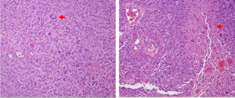
Figure 1:
On November 12, 2019, the Institute of Neurology and Neurosurgery for neurosurgical procedure (November 13, 2019) where it was performed
Left Temporo-Parietal Craniotomy, during the transoperative period a frozen biopsy was performed where slight nuclear pleomorphism in the brain white matter was observed, there was no mitoses or newly formed vessels, K167 with marked nuclei less than 1%, concluding a histopathological diagnosis of reactive gliosis secondary to a low-grade tumor.
A CT imaging study of the Post Cranial Nervous System is performed Operative (November 17, 2019) observing image of bone continuity solution in the left parietal region related to the craniotomy area performed during the surgery, noting little residual edema in the surgical bed, which is visualized as a hypodense image in the left parietal cortico-subcortical region. No hematomas are observed in the bed, and the presence of discrete bilateral frontal pneumocephalus is observed. Midline structures are intact, no displacement of the same, ventricular system and basal cisterns are present and functional.
The patient presented adequate clinical evolution, the wound showed no signs of local infection, and was discharged on November 13, 2019.
On November 17, 2019, the patient presented a seizure, with focal onset, which later became generalized, without loss of consciousness, which is why the anticonvulsant dose of Phenytoin was adjusted to 250 mg every 8 hours, anti- edema measures were started with 20% Mannitol IV for 5 days and Betamethasone (4 mg ampoule) every 8 hours, the patient responded to said treatment obtaining an adequate recovery.
During this hospital stay, a Simple Cranial CT scan was performed (November 22, 2019) were, due to the resolution of the post-surgical cerebral edema, a residual tumor lesion could be seen. However, it was decided to discharge the patient with clinical and imaging follow-up by the neurosurgery outpatient clinic, and the procedure was started to complete the treatment with Radiosurgery, which was arranged to be performed outside the country, but due to the epidemiological situation in the country and the world (Pandemic - COVID 19) this process was not possible to carry out.
In November 2021, the patient returned to our institution for consultation due to motor dysphasia, right hemiparesis, and recurrent headaches that did not respond to analgesic treatment.
A simple CT scan of the head was performed, which showed an increase in the size of the previously addressed tumor lesion accompanied by a cystic component, which compressed the ipsilateral ventricle and displaced midline structures. Given these imaging findings, it was decided to readmit on November 20, 2021, and the imaging study was expanded with contrast-enhanced brain MRI (November 23, 2021). Where a complex occupying lesion can be seen in the left frontoparietal region, its solid component occupies the periphery of the lesion, restricts diffusion, shows signs of angiogenesis and captures contrast.
The lesion measured 7.4 x 6.21 cm in axial section and is associated with a mass effect on neighboring structures. It is taken to the operating room with the objective of draining the cystic component of the lesion, which was carried out on November 26, 2021, where 60 ml of a yellow-greenish liquid with an oily consistency was extracted. With cytochemical: Color: Yellow, Appearance: Transparent, Pandi +++, Glycemia: 6.2 ml, Proteins: 2.08 ml.
A simple CT scan of the head was performed after the procedure (November 26, 2021), documenting the expected post-surgical changes in the recent left temporo-parietal region, signs of residual tumor lesion, and no hemorrhagic or acute ischemic signs.
After this procedure, the patient presented a favorable clinical evolution with evident improvement of dysphagia, adequate articulation of words, still maintained paresis of the right upper limb with a muscle strength of 3/5 distal and distal and was discharged from the hospital on November 29, 2021.
On December 17, 2021, the patient began with the same condition previously presented. A simple head CT scan was performed, which documented the presence of the cystic portion treated less than a month ago again. For this reason, she was admitted again.
Operated on December 20, 2021 Puncture of the cystic portion of the Right Parieto-Temporal lesion, evacuation and placement of intracystic Ommaya reservoir (45 ml of yellow pressurized fluid was drained).
The next day the patient is discharged.
On January 5, 2022, the patient was seen due to temperature increases of 38.5 °C. The reservoir was punctured, draining 28 ml and a medical discharge was decided. On January 8, 2022, the patient presented a focal onset seizure, which later became generalized. Convulsive management was adjusted. A simple head CT scan was performed (Figure 2) which documented an extensive, already known, occupying lesion with a drainage system in place, mass effect on the parenchyma and ventricular system.
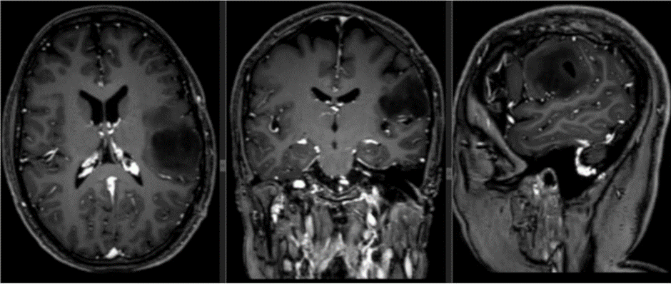
Figure 2
5 days later (January 10, 2022) he returned again, the reservoir was punctured but fluid extraction was not possible. Therefore, he was admitted for study.
A CEREBRAL MRI imaging study was performed (January 22, 2022) (Figure 3) surgical area occupied by a cystic portion of a dedifferentiated high- grade Astrocytoma tumor requiring puncture and evacuation of the cystic portion with a late control imaging study with tumor recurrence.
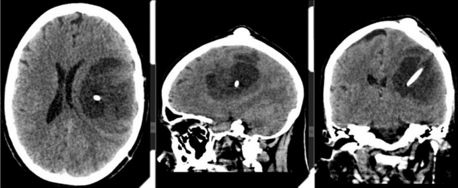
Figure 3
An electroencephalogram was performed (January 12, 2022) in which signs of intercritical focal cortical irritation of the left temporo-parietal regions of moderate intensity, signs of cortical dysfunction of the left cerebellar hemisphere of slight intensity were documented.
On January 18, 2022, the same previous surgery was performed at the left parieto-temporal level and the maximum safe macroscopic resection of the lesion was performed with a biopsy result (January 20, 2022): In the sample examined, hypercellularity and pleomorphism were observed.
cellular with the presence of tumor gemistocytic, small and other elongated cells. High number of atypical mitoses of endothelial hyperplasia with a glomeruloid appearance, microscopic areas of necrosis in palisade. Diagnosis: IDH MUTATED GLIOBLASTOMA. WHO GRADE IV.
The following day, January 20, 2022, a Simple Cranial CT scan (Figure 4) was performed for post-surgical control, observing recent post-surgical changes associated with a region of left cerebral hemispheric edema.
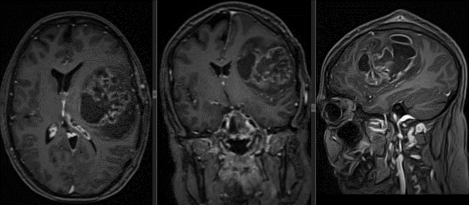
Figure 4
High-field imaging control was performed with Contrast-Enhanced Brain MRI (January 21, 2022) and a known heterogeneous left frontoparietal lesion was documented with significant mass effect on neighboring structures. After the administration of contrast, the lesion enhances in a non-homogeneous manner associated with enhancement of the ipsilateral ependyma as well as signs of diffusion restriction.
Patient with adequate clinical evolution, discharged on January 25, 2022 with referral by the Oncology service to start Radiation treatment completing 33 sessions of 1.8 GY which culminated on May 22, 2022 and Temozolamide concurrent with radiotherapy at a dose of 75 mg/m2 daily for 49 days and subsequently Adjuvant at a dose of 170 mg/m2 daily for 5 days every 28 days
On June 6, 2022, they consulted the institute of neurology and neurosurgery due to a clinical picture of one month of evolution, given by progressive decrease in muscle strength in the right side of the body, inability to express language, intermittent headache of mild to moderate intensity, occasional photophobia, reasons for which it was decided to admit them. A simple cranial CT study was performed (June 6, 2022) (Figure 5) observing post-surgical changes, an occupying lesion in the left fronto-parietal region measuring approximately 6.3 x 9.7 cm, heterogeneous with predominantly fluid (15 HU). This lesion exerts a mass effect on the ventricular system and neighboring parenchyma. IDX: Glioma.
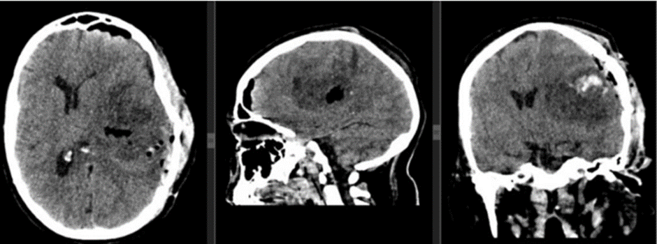
Figure 5
Given these findings, it was decided to move the neurosurgical procedure to a new date (June 22, 2022), the injury from previous surgery was addressed, immediately documenting the leakage of cerebrospinal fluid upon opening the dura mater, and the remaining tumor was addressed in the BIOPSY result. (Figure 12)
Pleomorphic tumor cells, high mitotic index, areas of ischemic necrosis, and collagenized vessel walls are observed. Tumor cells are dispersed away from the tumor core. A diagnosis of IDH MUTATED GLIOBLASTOMA, RECURRENT, WHO GRADE 4 is concluded (IDH: Performed at the Hermanos Amejeiras Hospital)
Simple Cranial CT scan is taken (June 23, 2022) (Figure 6) Recent postoperative changes in the left frontotemporal region. No signs of hydrocephalus. No signs of ischemic injury or acute hemorrhage. The edema in the surgical bed is striking, with blood in its interior, which seems to extend to the inferior longitudinal sinus. The bone flap is dislocated.
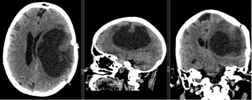
Figure 6
Simple Cranial CT (June 27, 2022) (Figure 7)
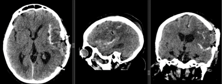
Figure 7
Postoperative changes in the left frontoparietal region are not recent, comparatively cerebral edema has increased in the left cerebral hemisphere and it is striking that the areas of bleeding in the surgical bed are larger. as well as herniation of the brain and an increase in the volume of the epicranial soft tissues. It is striking that the bone flap is more dislocated than in the previous study.
On July 14, 2022, the patient had a transcalvarian hernia confirmed by a Simple Cranial CT scan (Figure 8) accompanied by dehiscence of the surgical wound, for which she was taken to the operating room for repair, with respective imaging control (Figure 9). On August 22, 2022, an electroencephalogram was performed to adjust anticonvulsant treatment, a 19-channel EEG was performed, and the patient was in a functional state of wakefulness. Continuous baseline activity was observed, with preserved frequency and amplitude gradients. Globally, better modulated alpha activity of greater amplitude was recorded in the right hemisphere, alternating with diffuse global polymorphic theta activity, which was not blocked by eye opening.
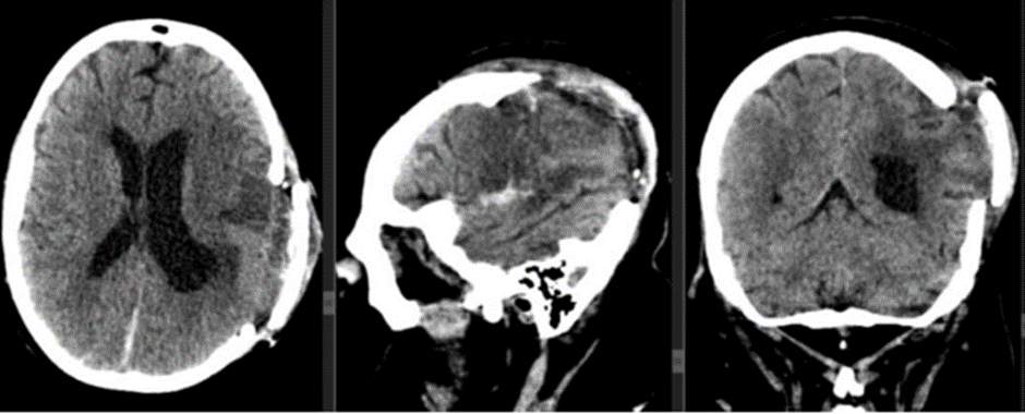
Figure 8
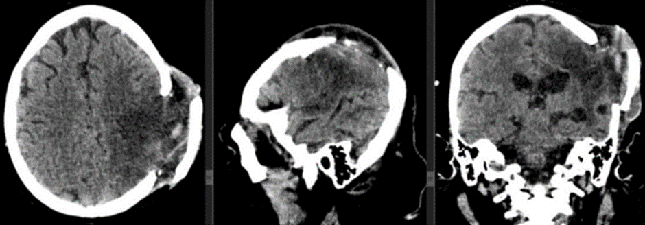
Figure 9
Focal interictal paroxysmal activity is recorded in the central-parietal region of the right hemisphere, of the type of slow angular waves that appear as very few isolated elements in the recording. Abnormal EEG. Signs of cortical irritation in the central-parietal region of the right hemisphere, of slight intensity
Patient is evolving satisfactorily with discharge hospitalized on September 20, 2022.
On October 4th, he starts with adjuvant treatment: RT + HeberFERON (currently 7.0 MUI: 2 bulbs - Tuesday and Friday) so far 2 years and 5 months have been completed (29 months). In February 2023, a Brain MRI is taken. (Figure 10) evolutionary where tumor control is observed.
The patient is currently at Miami Neuroscience Center at Larkin Community Hospital Inc. Dr. Aizik L. Wolf where imaging control was performed 19 months after starting Heberferon (Figure 11).
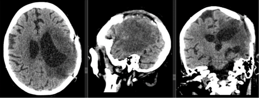
Figure 10
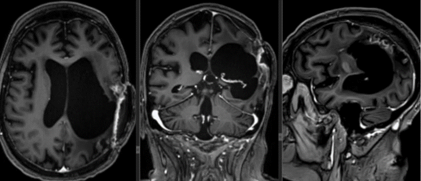
Figure 11
(Figure 12). Remarkable nuclear pleomorphism and giant cell characteristics along with pseudopalisade necrosis.
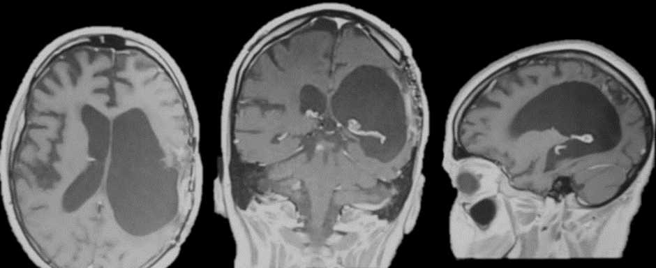
Figure 12
The second case is a 63-year-old male patient with a history of benign prostatic hyperplasia who underwent surgery in 2018. He debuted in January 2022 with associated gait instability, associated with intense, oppressive, holocranial headache, predominantly in the morning with little response to taking analgesics. No positive findings were found in the neurological physical examination.
In the emergency room, a simple head CT scan was performed, which showed mixed density images in the right fronto-parieto-temporal region, producing a mass effect on the ipsilateral lateral ventricle, as well as displacement of midline structures, surrounded by abundant perilesional edema.
An electroencephalogram was performed, which was reported as normal.
A brain MRI study shows a right sylvian occupying lesion surrounded by vasogenic edema with mass effect on neighboring structures with focal areas of diffusion restriction, signs of angiogenesis and calcification nuclei inside.
After administration of contrast, the walls enhance intensely, appearing irregular and thickened (Figure 13).
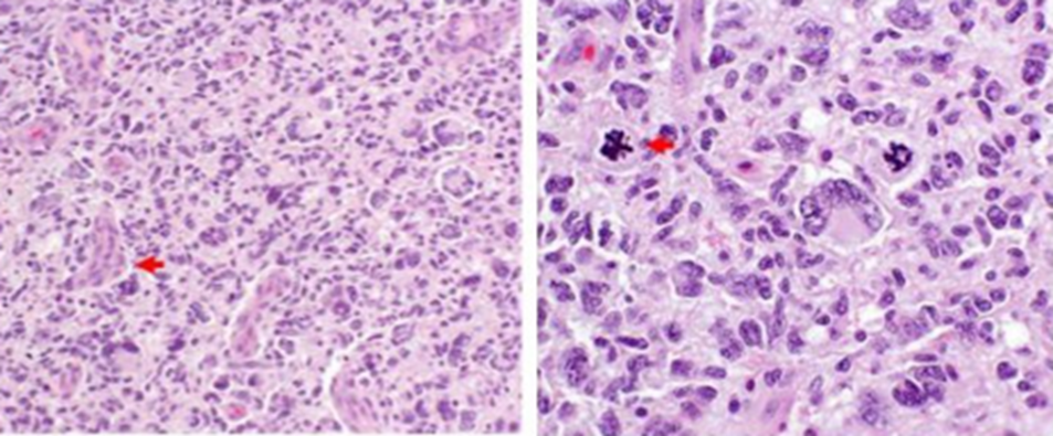
Figure 13
The spectroscopy sequence shows a high lipid peak. The lesion described measures 2.3 x 3 cm, which is related to Glioblastoma. Due to these findings, at the end of February 2022 he was taken to surgery where a right parieto-temporal craniotomy was performed with total fluoroguided tumor excision.
The biopsy study confirmed the diagnosis of Glioblastoma NOS Grade IV WHO, and the microscopic study of the sample showed fragments of cortex with the presence of red neurons and neuropil edema. Abundant areas of ischemic necrosis, vessels with thickened walls of glomeruloid appearance, newly formed vessels, a few small tumor cells with pyknocytic nuclei, and other large pleomorphic cells with atypical mitoses were observed. (Figure 14).
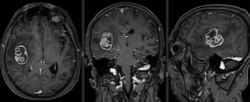
Figure 14
One week after surgery, he was discharged with adjuvant treatment: RT + Temozolamide alone as monotherapy (6 cycles) + HeberFERON. In February 2023, a progressive brain MRI was taken where tumor control was observed (Figure 15).
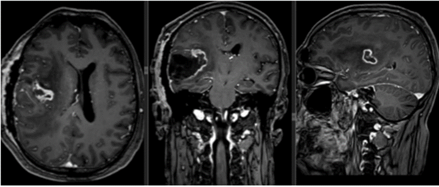
Figure 15
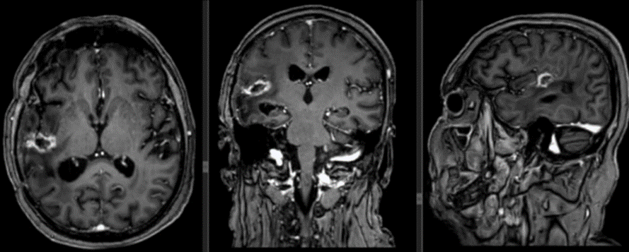
Figure 16
Clearly Auctoresonline and particularly Psychology and Mental Health Care Journal is dedicated to improving health care services for individuals and populations. The editorial boards' ability to efficiently recognize and share the global importance of health literacy with a variety of stakeholders. Auctoresonline publishing platform can be used to facilitate of optimal client-based services and should be added to health care professionals' repertoire of evidence-based health care resources.

Journal of Clinical Cardiology and Cardiovascular Intervention The submission and review process was adequate. However I think that the publication total value should have been enlightened in early fases. Thank you for all.

Journal of Women Health Care and Issues By the present mail, I want to say thank to you and tour colleagues for facilitating my published article. Specially thank you for the peer review process, support from the editorial office. I appreciate positively the quality of your journal.
Journal of Clinical Research and Reports I would be very delighted to submit my testimonial regarding the reviewer board and the editorial office. The reviewer board were accurate and helpful regarding any modifications for my manuscript. And the editorial office were very helpful and supportive in contacting and monitoring with any update and offering help. It was my pleasure to contribute with your promising Journal and I am looking forward for more collaboration.

We would like to thank the Journal of Thoracic Disease and Cardiothoracic Surgery because of the services they provided us for our articles. The peer-review process was done in a very excellent time manner, and the opinions of the reviewers helped us to improve our manuscript further. The editorial office had an outstanding correspondence with us and guided us in many ways. During a hard time of the pandemic that is affecting every one of us tremendously, the editorial office helped us make everything easier for publishing scientific work. Hope for a more scientific relationship with your Journal.

The peer-review process which consisted high quality queries on the paper. I did answer six reviewers’ questions and comments before the paper was accepted. The support from the editorial office is excellent.

Journal of Neuroscience and Neurological Surgery. I had the experience of publishing a research article recently. The whole process was simple from submission to publication. The reviewers made specific and valuable recommendations and corrections that improved the quality of my publication. I strongly recommend this Journal.

Dr. Katarzyna Byczkowska My testimonial covering: "The peer review process is quick and effective. The support from the editorial office is very professional and friendly. Quality of the Clinical Cardiology and Cardiovascular Interventions is scientific and publishes ground-breaking research on cardiology that is useful for other professionals in the field.

Thank you most sincerely, with regard to the support you have given in relation to the reviewing process and the processing of my article entitled "Large Cell Neuroendocrine Carcinoma of The Prostate Gland: A Review and Update" for publication in your esteemed Journal, Journal of Cancer Research and Cellular Therapeutics". The editorial team has been very supportive.

Testimony of Journal of Clinical Otorhinolaryngology: work with your Reviews has been a educational and constructive experience. The editorial office were very helpful and supportive. It was a pleasure to contribute to your Journal.

Dr. Bernard Terkimbi Utoo, I am happy to publish my scientific work in Journal of Women Health Care and Issues (JWHCI). The manuscript submission was seamless and peer review process was top notch. I was amazed that 4 reviewers worked on the manuscript which made it a highly technical, standard and excellent quality paper. I appreciate the format and consideration for the APC as well as the speed of publication. It is my pleasure to continue with this scientific relationship with the esteem JWHCI.

This is an acknowledgment for peer reviewers, editorial board of Journal of Clinical Research and Reports. They show a lot of consideration for us as publishers for our research article “Evaluation of the different factors associated with side effects of COVID-19 vaccination on medical students, Mutah university, Al-Karak, Jordan”, in a very professional and easy way. This journal is one of outstanding medical journal.
Dear Hao Jiang, to Journal of Nutrition and Food Processing We greatly appreciate the efficient, professional and rapid processing of our paper by your team. If there is anything else we should do, please do not hesitate to let us know. On behalf of my co-authors, we would like to express our great appreciation to editor and reviewers.

As an author who has recently published in the journal "Brain and Neurological Disorders". I am delighted to provide a testimonial on the peer review process, editorial office support, and the overall quality of the journal. The peer review process at Brain and Neurological Disorders is rigorous and meticulous, ensuring that only high-quality, evidence-based research is published. The reviewers are experts in their fields, and their comments and suggestions were constructive and helped improve the quality of my manuscript. The review process was timely and efficient, with clear communication from the editorial office at each stage. The support from the editorial office was exceptional throughout the entire process. The editorial staff was responsive, professional, and always willing to help. They provided valuable guidance on formatting, structure, and ethical considerations, making the submission process seamless. Moreover, they kept me informed about the status of my manuscript and provided timely updates, which made the process less stressful. The journal Brain and Neurological Disorders is of the highest quality, with a strong focus on publishing cutting-edge research in the field of neurology. The articles published in this journal are well-researched, rigorously peer-reviewed, and written by experts in the field. The journal maintains high standards, ensuring that readers are provided with the most up-to-date and reliable information on brain and neurological disorders. In conclusion, I had a wonderful experience publishing in Brain and Neurological Disorders. The peer review process was thorough, the editorial office provided exceptional support, and the journal's quality is second to none. I would highly recommend this journal to any researcher working in the field of neurology and brain disorders.

Dear Agrippa Hilda, Journal of Neuroscience and Neurological Surgery, Editorial Coordinator, I trust this message finds you well. I want to extend my appreciation for considering my article for publication in your esteemed journal. I am pleased to provide a testimonial regarding the peer review process and the support received from your editorial office. The peer review process for my paper was carried out in a highly professional and thorough manner. The feedback and comments provided by the authors were constructive and very useful in improving the quality of the manuscript. This rigorous assessment process undoubtedly contributes to the high standards maintained by your journal.

International Journal of Clinical Case Reports and Reviews. I strongly recommend to consider submitting your work to this high-quality journal. The support and availability of the Editorial staff is outstanding and the review process was both efficient and rigorous.

Thank you very much for publishing my Research Article titled “Comparing Treatment Outcome Of Allergic Rhinitis Patients After Using Fluticasone Nasal Spray And Nasal Douching" in the Journal of Clinical Otorhinolaryngology. As Medical Professionals we are immensely benefited from study of various informative Articles and Papers published in this high quality Journal. I look forward to enriching my knowledge by regular study of the Journal and contribute my future work in the field of ENT through the Journal for use by the medical fraternity. The support from the Editorial office was excellent and very prompt. I also welcome the comments received from the readers of my Research Article.

Dear Erica Kelsey, Editorial Coordinator of Cancer Research and Cellular Therapeutics Our team is very satisfied with the processing of our paper by your journal. That was fast, efficient, rigorous, but without unnecessary complications. We appreciated the very short time between the submission of the paper and its publication on line on your site.

I am very glad to say that the peer review process is very successful and fast and support from the Editorial Office. Therefore, I would like to continue our scientific relationship for a long time. And I especially thank you for your kindly attention towards my article. Have a good day!

"We recently published an article entitled “Influence of beta-Cyclodextrins upon the Degradation of Carbofuran Derivatives under Alkaline Conditions" in the Journal of “Pesticides and Biofertilizers” to show that the cyclodextrins protect the carbamates increasing their half-life time in the presence of basic conditions This will be very helpful to understand carbofuran behaviour in the analytical, agro-environmental and food areas. We greatly appreciated the interaction with the editor and the editorial team; we were particularly well accompanied during the course of the revision process, since all various steps towards publication were short and without delay".

I would like to express my gratitude towards you process of article review and submission. I found this to be very fair and expedient. Your follow up has been excellent. I have many publications in national and international journal and your process has been one of the best so far. Keep up the great work.

We are grateful for this opportunity to provide a glowing recommendation to the Journal of Psychiatry and Psychotherapy. We found that the editorial team were very supportive, helpful, kept us abreast of timelines and over all very professional in nature. The peer review process was rigorous, efficient and constructive that really enhanced our article submission. The experience with this journal remains one of our best ever and we look forward to providing future submissions in the near future.

I am very pleased to serve as EBM of the journal, I hope many years of my experience in stem cells can help the journal from one way or another. As we know, stem cells hold great potential for regenerative medicine, which are mostly used to promote the repair response of diseased, dysfunctional or injured tissue using stem cells or their derivatives. I think Stem Cell Research and Therapeutics International is a great platform to publish and share the understanding towards the biology and translational or clinical application of stem cells.

I would like to give my testimony in the support I have got by the peer review process and to support the editorial office where they were of asset to support young author like me to be encouraged to publish their work in your respected journal and globalize and share knowledge across the globe. I really give my great gratitude to your journal and the peer review including the editorial office.

I am delighted to publish our manuscript entitled "A Perspective on Cocaine Induced Stroke - Its Mechanisms and Management" in the Journal of Neuroscience and Neurological Surgery. The peer review process, support from the editorial office, and quality of the journal are excellent. The manuscripts published are of high quality and of excellent scientific value. I recommend this journal very much to colleagues.

Dr.Tania Muñoz, My experience as researcher and author of a review article in The Journal Clinical Cardiology and Interventions has been very enriching and stimulating. The editorial team is excellent, performs its work with absolute responsibility and delivery. They are proactive, dynamic and receptive to all proposals. Supporting at all times the vast universe of authors who choose them as an option for publication. The team of review specialists, members of the editorial board, are brilliant professionals, with remarkable performance in medical research and scientific methodology. Together they form a frontline team that consolidates the JCCI as a magnificent option for the publication and review of high-level medical articles and broad collective interest. I am honored to be able to share my review article and open to receive all your comments.

“The peer review process of JPMHC is quick and effective. Authors are benefited by good and professional reviewers with huge experience in the field of psychology and mental health. The support from the editorial office is very professional. People to contact to are friendly and happy to help and assist any query authors might have. Quality of the Journal is scientific and publishes ground-breaking research on mental health that is useful for other professionals in the field”.

Dear editorial department: On behalf of our team, I hereby certify the reliability and superiority of the International Journal of Clinical Case Reports and Reviews in the peer review process, editorial support, and journal quality. Firstly, the peer review process of the International Journal of Clinical Case Reports and Reviews is rigorous, fair, transparent, fast, and of high quality. The editorial department invites experts from relevant fields as anonymous reviewers to review all submitted manuscripts. These experts have rich academic backgrounds and experience, and can accurately evaluate the academic quality, originality, and suitability of manuscripts. The editorial department is committed to ensuring the rigor of the peer review process, while also making every effort to ensure a fast review cycle to meet the needs of authors and the academic community. Secondly, the editorial team of the International Journal of Clinical Case Reports and Reviews is composed of a group of senior scholars and professionals with rich experience and professional knowledge in related fields. The editorial department is committed to assisting authors in improving their manuscripts, ensuring their academic accuracy, clarity, and completeness. Editors actively collaborate with authors, providing useful suggestions and feedback to promote the improvement and development of the manuscript. We believe that the support of the editorial department is one of the key factors in ensuring the quality of the journal. Finally, the International Journal of Clinical Case Reports and Reviews is renowned for its high- quality articles and strict academic standards. The editorial department is committed to publishing innovative and academically valuable research results to promote the development and progress of related fields. The International Journal of Clinical Case Reports and Reviews is reasonably priced and ensures excellent service and quality ratio, allowing authors to obtain high-level academic publishing opportunities in an affordable manner. I hereby solemnly declare that the International Journal of Clinical Case Reports and Reviews has a high level of credibility and superiority in terms of peer review process, editorial support, reasonable fees, and journal quality. Sincerely, Rui Tao.

Clinical Cardiology and Cardiovascular Interventions I testity the covering of the peer review process, support from the editorial office, and quality of the journal.

Clinical Cardiology and Cardiovascular Interventions, we deeply appreciate the interest shown in our work and its publication. It has been a true pleasure to collaborate with you. The peer review process, as well as the support provided by the editorial office, have been exceptional, and the quality of the journal is very high, which was a determining factor in our decision to publish with you.
The peer reviewers process is quick and effective, the supports from editorial office is excellent, the quality of journal is high. I would like to collabroate with Internatioanl journal of Clinical Case Reports and Reviews journal clinically in the future time.

Clinical Cardiology and Cardiovascular Interventions, I would like to express my sincerest gratitude for the trust placed in our team for the publication in your journal. It has been a true pleasure to collaborate with you on this project. I am pleased to inform you that both the peer review process and the attention from the editorial coordination have been excellent. Your team has worked with dedication and professionalism to ensure that your publication meets the highest standards of quality. We are confident that this collaboration will result in mutual success, and we are eager to see the fruits of this shared effort.

Dear Dr. Jessica Magne, Editorial Coordinator 0f Clinical Cardiology and Cardiovascular Interventions, I hope this message finds you well. I want to express my utmost gratitude for your excellent work and for the dedication and speed in the publication process of my article titled "Navigating Innovation: Qualitative Insights on Using Technology for Health Education in Acute Coronary Syndrome Patients." I am very satisfied with the peer review process, the support from the editorial office, and the quality of the journal. I hope we can maintain our scientific relationship in the long term.
Dear Monica Gissare, - Editorial Coordinator of Nutrition and Food Processing. ¨My testimony with you is truly professional, with a positive response regarding the follow-up of the article and its review, you took into account my qualities and the importance of the topic¨.

Dear Dr. Jessica Magne, Editorial Coordinator 0f Clinical Cardiology and Cardiovascular Interventions, The review process for the article “The Handling of Anti-aggregants and Anticoagulants in the Oncologic Heart Patient Submitted to Surgery” was extremely rigorous and detailed. From the initial submission to the final acceptance, the editorial team at the “Journal of Clinical Cardiology and Cardiovascular Interventions” demonstrated a high level of professionalism and dedication. The reviewers provided constructive and detailed feedback, which was essential for improving the quality of our work. Communication was always clear and efficient, ensuring that all our questions were promptly addressed. The quality of the “Journal of Clinical Cardiology and Cardiovascular Interventions” is undeniable. It is a peer-reviewed, open-access publication dedicated exclusively to disseminating high-quality research in the field of clinical cardiology and cardiovascular interventions. The journal's impact factor is currently under evaluation, and it is indexed in reputable databases, which further reinforces its credibility and relevance in the scientific field. I highly recommend this journal to researchers looking for a reputable platform to publish their studies.

Dear Editorial Coordinator of the Journal of Nutrition and Food Processing! "I would like to thank the Journal of Nutrition and Food Processing for including and publishing my article. The peer review process was very quick, movement and precise. The Editorial Board has done an extremely conscientious job with much help, valuable comments and advices. I find the journal very valuable from a professional point of view, thank you very much for allowing me to be part of it and I would like to participate in the future!”

Dealing with The Journal of Neurology and Neurological Surgery was very smooth and comprehensive. The office staff took time to address my needs and the response from editors and the office was prompt and fair. I certainly hope to publish with this journal again.Their professionalism is apparent and more than satisfactory. Susan Weiner

My Testimonial Covering as fellowing: Lin-Show Chin. The peer reviewers process is quick and effective, the supports from editorial office is excellent, the quality of journal is high. I would like to collabroate with Internatioanl journal of Clinical Case Reports and Reviews.

My experience publishing in Psychology and Mental Health Care was exceptional. The peer review process was rigorous and constructive, with reviewers providing valuable insights that helped enhance the quality of our work. The editorial team was highly supportive and responsive, making the submission process smooth and efficient. The journal's commitment to high standards and academic rigor makes it a respected platform for quality research. I am grateful for the opportunity to publish in such a reputable journal.
My experience publishing in International Journal of Clinical Case Reports and Reviews was exceptional. I Come forth to Provide a Testimonial Covering the Peer Review Process and the editorial office for the Professional and Impartial Evaluation of the Manuscript.

I would like to offer my testimony in the support. I have received through the peer review process and support the editorial office where they are to support young authors like me, encourage them to publish their work in your esteemed journals, and globalize and share knowledge globally. I really appreciate your journal, peer review, and editorial office.
Dear Agrippa Hilda- Editorial Coordinator of Journal of Neuroscience and Neurological Surgery, "The peer review process was very quick and of high quality, which can also be seen in the articles in the journal. The collaboration with the editorial office was very good."
