AUCTORES
Globalize your Research

Research Article | DOI: https://doi.org/10.31579/2578-8965/269
1 Master's degree in Medical Education. First- degree specialist in Comprehensive General Medicine and Histology. Assistant Professor. University of Medical Sciences Holguín. Mariana Grajales Coello Faculty of Medical Sciences. Department of Basic Sciences. Holguín, Cuba.
2 Doctor of Science in Medical Education. Master of Science in Medical Education. Second-Degree Specialist in Histology. Second-Degree Specialist in Health Administration. Full Professor. Senior Researcher. University of Medical Sciences. Mariana Grajales Coello Faculty of Medical Sciences. Department of Basic Sciences. Holguín, Cuba.
3 Master's Degree in Medical Education. First Degree Specialist in Comprehensive General Medicine and Histology. Assistant Professor. University of Medical Sciences Holguín. Mariana Grajales Coello Faculty of Medical Sciences. Department of Basic Sciences. Holguín, Cuba.
4 Master's Degree in Comprehensive Women's Care. Master's Degree in Medical Education. Second-Degree Specialist in Comprehensive General Medicine. First-Degree Specialist in Histology. Assistant Professor. University of Medical Sciences. Mariana Grajales Coello Faculty of Medical Sciences. Department of Basic Sciences. Holguín, Cuba.
*Corresponding Author: Dunia Yailin Macareño, Master's degree in Medical Education. First- degree specialist in Comprehensive General Medicine and Histology. Assistant Professor. University of Medical Sciences Holguín. Mariana Grajales Coello Faculty of Medical Sciences. Depart
Citation: Macareño Avila DY, Díaz Rojas PA, Doralny P. Marrero, Leticia M. Caballero, (2025), Morphometric Analysis of Fibroblast Nuclei in Women's Breast Stroma from 60 Years, J. Obstetrics Gynecology and Reproductive Sciences, 9(4) DOI:10.31579/2578-8965/269
Copyright: © 2025, Dunia Yailin Macareño. This is an open-access article distributed under the terms of The Creative Commons Attribution License, which permits unrestricted use, distribution, and reproduction in any medium, provided the original author and source are credited.
Received: 09 May 2025 | Accepted: 19 May 2025 | Published: 29 May 2025
Keywords: Cell core; fibroblasts; human mammary glands; aging; women
Introduction: Aging is a physiological process that reaches all organs and produces histological alterations characteristic in them. The female mammary glands undergo changes because of the passage of time. The changes that the populations of fibroblasts have mainly regarding their number linked to aging have been studied but there are few studies linked to nuclear changes in aging fibroblasts and even less those belonging to the stroma of the mammary glands of older adults.
Objectives: Characterize the perimeter area volume and shape factor in fibroblasts nuclei from healthy mammary glands in women aged 60 years and older.
Methods: To characterize the healthy mammary glands in 60 -year -old women and a serial study in killed women who did not have benign or malignant lesions of the organ was carried out. All examined by the Department of Pathological Anatomy of the Vladimir Ilich Lenin Provincial Hospital in Holguín in the period from September 2018 to September 2019. For better assessment, the sample of studies was divided into two age groups from 60 to 75 years old and over 75 years.
Results: Both the area and volume perimeter and the form factor of the nuclei of the fibroblasts of the breast stroma are lower in women over 75 years
Conclusions: The size and shape of the fibroblast nuclei in the healthy mammary glands are affected with age being smaller and elongated in women over 75 years old
Aging, or senescence, is the set of morphological and physiological changes that occur as a result of the effects of time on living beings. It is an inevitable, irreversible, and complex biological reality resulting from complex processes of accumulated cellular and molecular damage.
In humans, aging is a process that begins at conception and develops throughout life. Biologically, all vital organs begin to lose functionality as age advances. Thus, changes in this process have been found in all cells, tissues, and organs of the body and affect the functioning of all bodily systems. This is a physiological process that affects all organs and produces characteristic histological changes in them.
Female mammary glands undergo changes with the passage of time. Some authors [3,4] describe these changes in the breasts of older women quantitatively using morphometry. These studies focus primarily on histological changes in the breast parenchyma.
The literature describes an increase in stromal structures and a decrease in parenchyma during the involution of female mammary glands. The mammary stroma is composed of connective tissue, which changes as a woman ages, particularly during postmenopause. [5].
The fibroblast is the predominant cell in the body's connective tissues. It is a dynamic cell that performs local tissue functions and functions within the immune system. When evaluated in culture, it has been interestingly found that fibroblasts are not homogeneous, but rather differ in morphology and function depending on their location. [6,7]
Several authors have studied the changes in fibroblast populations, primarily related to their number, linked to skin aging [1, 8] and periodontal disorders. [9, 10] However, there are few studies related to nuclear changes in aged fibroblasts, and even fewer are those in the stroma of the mammary glands of older women.
Not all the possibilities offered by morphometric techniques are exploited for this purpose, so we decided to carry out a morphometric study with the aim of analyzing the behavior of the nuclear indicators of fibroblasts in healthy mammary glands of women aged 60 years and older, in order to establish reference patterns in non-pathological breast tissue, knowledge that will be useful in teaching and research.
A case series study was conducted in the female population aged 60 years and older in the province of Holguín.
The study universe consisted of deceased women of these ages, who were examined by the Department of Pathological Anatomy of the VILenin Provincial University Hospital in the period from September 2018 to September 2019.
A purposive, non-probability sample of 14 women with no history of mammary gland disease confirmed by examination was selected. Those with a history of benign or malignant mammary gland disease and/or those confirmed by autopsy were excluded.
To analyze the behavior of the indicators, the study sample was divided into two age groups: 60 to 75 years and those over 75 years.
A fragment of breast tissue corresponding to the upper inner quadrant was taken from the inside of each woman studied, given its easy availability and to respect the ethical aspects of the research.
Each tissue fragment was fixed in 10% formalin and embedded using the classic paraffin technique. Histological sections were 10 micrometers thick and stained with hematoxylin and eosin (H&E).
The histological study of the mammary gland was performed by observing the images in a Chinese Motic microscope, model BA-210 with a 3 megapixel camera from the Motic company, with a 100X objective lens and a 10X eyepiece lens. The images were downloaded to a Dell computer, model Optiplex 7010, to which the microscope capture system was coupled. The total magnification (At) of observation of the images captured on the computer was calculated using the formula:
A t = Objective Magnification x Reducer Lens Magnification x Screen Length / Digital Camera Sensor
In this way, the total magnification with which the histological images were processed when captured on the computer used for this purpose was 2125 X.
To calculate the shape factor, area, perimeter and nuclear volume of stromal fibroblasts from the mammary glands, ImageJ software, version 1.49p from the National Institutes of Health, USA, 2015, was used. (Figure 1)

Figure 1: Presentation of the ImageJ 1.49p application.
Connective tissue nuclei that matched the nuclear histological characteristics of fibroblasts were selected for study, with clearly visible boundaries and no overlap. With this selection, measurements were taken on a total of 254 nuclei.
The shape factor was obtained using the Shape Descriptor option. The permissible outer edge of the cell nuclei was contoured using the ImageJ system's freehand algorithm (Figure 2). The system returns the circularity or shape factor value, using the perimeter and area indicators automatically calculated by the application.
ImageJ freehand contouring option of the admissible outer edge of cell nuclei (Figure 2).
The nuclear volume was obtained with the Fit Ellipse option, the admissible external edge of the cell nuclei was contoured freehand using the ImageJ system (Figure 2).
The system automatically returns the largest and smallest diameters. These values allow for the calculation of the nuclear volume of epithelial cells. The largest and smallest diameter data were entered into a Microsoft Excel spreadsheet, and Palkovits' formula (Formula 1) was applied: [11]

Where:
A: Largest diameter.
B: Smaller diameter.
π: 3.1416
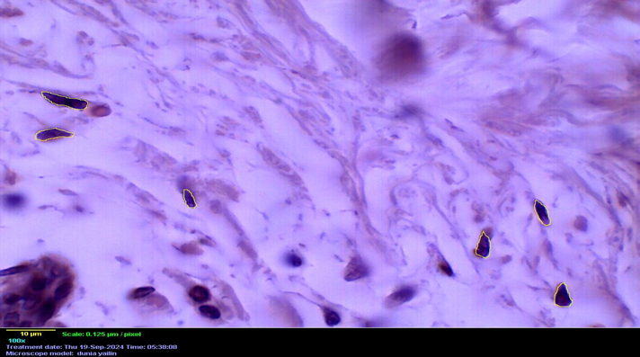
Figure 2: Microphotograph of a histological section of a mammary gland showing how the edges of the nuclei are delimited. Total magnification 2125X. H&E staining.
These indicators were analyzed separately according to the age groups established in the study. The data underwent a review process to avoid errors, omissions, and/or duplication of information. Extreme data, which were very far from the average values, were eliminated.
The results were processed using IBM SPSS Statistics 19v for Windows. Descriptive statistics were used to summarize the information: maximum and minimum values, arithmetic mean, standard deviation, and a normal distribution test for the different data series. Within the inferential statistics, a difference of means test was performed to compare the values obtained between different data groups. For the analysis, a 95% confidence interval was used, with a P ≤ 0.05 to assess statistical significance.
The ethical principles for medical research with data from human subjects of the World Medical Association Declaration of Helsinki were taken into account. and the WHO Guidelines for Research Ethics Committees, established by the Council for International Organizations of Medical Sciences (CIOMS) in 2002.
Area is a measure of the surface area, and in this case, it refers to the area of the cell nucleus. It is a two-dimensional unit whose value is obtained from a formula. This indicator allows us to determine the size of the cell nucleus on a surface. It allows us to interpret the size of the cell nucleus and the cell itself. This indicator is used to compare the state of nuclei under different conditions.
Volume is a scalar metric defined as the three-dimensional extension of a region of space. In the case of nuclear volume, it allows us to quantitatively define the three-dimensional extension of the cell nucleus.
When the mean-matching test was performed, significant differences were found between the two age groups studied. The results for the indicators that quantify the size of the nuclei (perimeter, area, and volume) were higher in the group of women aged 60 to 75. This means that the nuclei of the mammary stoma fibroblasts are smaller with age.
The nuclear shape factor or circularity index is the degree to which the shape of the nucleus in a cross-section approximates a perfect circle. The formula used to calculate this indicator relates the value of the nuclear area to the square of the nuclear perimeter, in a fraction from which it is inferred that the larger the nuclear perimeter, the closer the value is to one, indicating a perfect circle. If the perimeter value is low, the result tends to move away from one and approach zero, indicating an increase in the elongated shape. [11]
The results for the shape factor were similar, as the mean-matching test revealed differences in both age groups, with the lowest values found in the oldest group. This suggests that with age, the fibroblast nuclei in healthy mammary glands become more elongated or lengthened. These results are shown in Table 1.
| Indicators | GM * of women aged 60-75 MA ** -DS *** | GM of women over 75 years old MA-DS | Hypothesis testing |
| Number of cores | 127 | 127 | |
| Perimeter (µm) | 18,119–3,785 | 17.863-0.118 | p≥ 0.001 |
| Area ( μm | 14,440–4,570 | 12,660–4,031 | p <0.05 |
| Volume ( μm | 24,089–12,102 | 18,605–9,081 | p≥ 0.001 |
| Form factor (µm) | 0.563–0.123 | 0.504–0.118 | p ≥ 0.001 |
Table 1: Characterization of the behavior of nuclear indicators in fibroblasts of healthy mammary glands.
Morphometric and stereological parameters are tools that allow for a better understanding of pathological and physiological processes such as aging. Their effectiveness has been demonstrated, and they have allowed us to increase our knowledge of cellular and tissue structure, as well as the various physiological and pathological processes to which these elements are subject.
It has been demonstrated by different authors that the morphological analysis of the nucleus, including morphometric and stereological elements, can provide important information on the structure and morphology of cells and tissues and thus contribute to diagnostic and prognostic studies of lesions. Similarly, it is known that changes in the cell cycle or cellular metabolism, during different physiological states or pharmacological therapeutic interventions are accompanied by changes in nuclear architecture. [11]
Several authors have studied nuclear markers of different tissues linked to diseases such as neoplasia, [12, 13, 14] in experimental models [15, 16] and also in physiological processes such as aging. [2, 3, 8, 17].
Trasobares et al., [12] in their study on Basal Cell Carcinoma found that nuclear dimensions such as area and volume are larger compared to healthy skin. Similar results were reported by Toledo et al., [13] in Papillary Thyroid Carcinoma in contrast to Thyroid Nodule. These results reaffirm that most cancer cells undergo morphological changes, usually due to mutations in the corresponding genes, such as self-sufficiency in growth signals and insensitivity to growth inhibitory signals. [13].
In nuclear morphometric studies linked to aging, such as that carried out by Peña et al., [17] it was found that as age advances the perimeter, area and nuclear volume of keratinocytes decrease.
In a previous study by Macareño et al., 3 which morphometrically characterized the nuclei of epithelial cells from the ducts of healthy mammary glands from older women, it was found that the perimeter, area, and volume decreased with increasing age. Furthermore, it was found that circularity was not lost, although in the nuclei of the older group, the shape factor was smaller without becoming elongated.
Fibroblasts, as the fundamental cell of connective tissue and widely distributed throughout the body, have been the subject of several investigations. The most frequent ones relate to their multiple functions in dentistry [7, 10, 18] as well as in relation to ligament repair in sprains. [19, 20]
Research conducted by Simancas et al., [7,10] on gingival fibroblasts in young patients using qualitative studies, found that the size of the nuclei of these cells was voluminous and their shape was elongated and oval.
The literature [5,21] describes the qualitative changes that the breast stroma undergoes in older women. These changes include connective tissue atrophy, marked by a decrease in the number of fibroblasts and collagen fibers and the disappearance of elastic fibers. The intercellular substance appears to undergo hyaline degeneration. Furthermore, the radiodense fibrous stroma of young women is progressively replaced by radiolucent adipose tissue.
No research was found that addressed quantitative changes in nuclei in mammary stromal fibroblasts during aging. However, the behavior of the nuclear indicators studied by the authors is similar to a previous study by Macareño et al. 3 on nuclei of epithelial cells from healthy, aged mammary glands, with evidence of a decrease in nuclei with increasing age.
The use of morphometric techniques has allowed for the collection of quantitative data in studies on aging in certain locations of the body, such as the mammary glands, which makes the morphological characteristics that the passage of time leaves on our bodies more objective.
A significant decrease in the nuclear morphometric indicators of mammary stromal fibroblasts was observed in women over 75 years of age, reflected in lower values for perimeter, area, volume, and shape factor. These findings suggest that advanced aging is associated with morphological changes in fibroblasts, possibly related to processes of atrophy or cellular hypotrophy in breast tissue.
The results are consistent with the physiological involution of breast tissue in advanced age, where the reduction in size and alteration in nuclear shape could reflect a decrease in the metabolic and proliferative activity of these stromal cells. Therefore, the morphometric characterization of fibroblast nuclei provides a quantitative basis for better understanding the effects of aging on the mammary gland and establishes useful reference parameters for future comparative studies in physiological and pathological contexts.
Clearly Auctoresonline and particularly Psychology and Mental Health Care Journal is dedicated to improving health care services for individuals and populations. The editorial boards' ability to efficiently recognize and share the global importance of health literacy with a variety of stakeholders. Auctoresonline publishing platform can be used to facilitate of optimal client-based services and should be added to health care professionals' repertoire of evidence-based health care resources.


