

case report | DOI: https://doi.org/10.31579/2690-4861/1092
1Department of Cardiothoracic surgery, Tanta University Hospitals, Tanta, Egypt.
2Pediatric surgery unit, Department of General Surgery, Tanta University Hospitals, Tanta, Egypt.
*Corresponding Author: Mohammad Torky. MD, PhD, Faculty of Medicine, Department of Cardiothoracic surgery, Tanta University Hospitals, Tanta, Egypt.
Citation: Mohammad A. Torky, Ahmed M. Elsharaby, Sherif M. Negm, Samar A, Elrashedy, Alaa Basiouni, (2026), Large Iron Rod Thoraco-Abdominal Impalement in a Young Child: A Case Report, International Journal of Clinical Case Reports and Reviews, 35(3); DOI:10.31579/2690-4861/1092
Copyright: © 2026, Mohammad Torky. This is an open-access article distributed under the terms of the Creative Commons Attribution License, which permits unrestricted use, distribution, and reproduction in any medium, provided the original author and source are credited.
Received: 01 April 2026 | Accepted: 16 April 2026 | Published: 24 April 2026
Keywords: Type 2 diabetes mellitus; hyperglycemia; pathophysiology; hospitalization; hypoglycemia; pharmacology; corticosteroids; glibenclamide; insulins, hypoglycemia
Penetrating chest trauma is a major cause of mortality. Impalement injury happens when the penetrating object remains in situ at the time of presentation, patient’s clinical status and outcome is depending on the extent of the internal injuries.
We described a 15 years old child who was presented with a thoraco-abdominal impaled iron rod penetrating his body through the right lower costal margin at the posterior axillary line and exiting at the suprasternal notch. Preoperative computerized tomography with intravenous contrast showed the full course of the iron rod that was extracted under general anesthesia by cardiothoracic and abdominal surgeons.
Penetrating chest trauma is a major cause of mortality, as 10-15% only of these patients can reach the hospital alive and may need an emergent intervention due to vascular or visceral injury [1]. Thoracoabdominal penetrating trauma has been reported either by firearm or other injuries including road traffic accidents, stab wounds and impalements [2]. Impalement injury is produced by a large object penetrating the body cavities or extremities and remains in situ [3].
Almost all patients with impalement injuries needed surgical intervention and should be rapidly transferred to a tertiary care hospital, where a multidisciplinary team is available to get the optimal results [4]. In this article, we present a case of a 15 years old child with a thoracoabdominal impalement injury with a large iron rod.
A 15 years old child was presented to the emergency department of Tanta University Hospitals, Tanta, Egypt, after falling from a few meters’ height on an iron rod in a building under construction. The iron rod was sawed off in the trauma scene and the patient was transferred to the hospital with the rod impaling his body.
On admission, the patient was conscious, alert and hemodynamically stable. On examination, an iron rod of about 1 meter length and 1.5 cm in diameter was penetrating the right lower costal margin at the posterior axillary line, transfixing the thoracic cavity and exiting from the suprasternal notch (Figure.1).
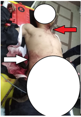
Figure 1: A photograph showing the inlet (white arrow) and exit (red arrow) of the iron rod on presentation.
There was neither palpable surgical emphysema nor fracture clicks.
Chest auscultation revealed a nearly equal bilateral air entry, audible heart sounds with no added sounds, no clinical signs of cardiac tamponade and radial pulse was equally palpable bilaterally.
Neurological and peripheral injuries were excluded.
CT-scan with intravenous contrast and bone reconstruction revealed mild right sided hydropneumothorax with the iron rod piercing the right 9th
intercostal space in the posterior axillary line, transfixing the liver, piercing the right copula of the diaphragm, passing anterior to the hilum of the right lung, on the right side of the pericardium then directing towards the anterior mediastinum and exiting from the suprasternal notch just behind the left sternoclavicular joint, with mild amount of free fluid in the abdomen and pelvis (Figure.2).
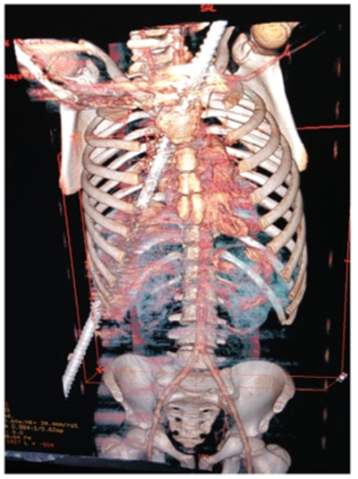
Figure 2: CT scan with bone reconstruction showing the course of the iron rod inside the body.
After discussion with abdominal surgeons considering the CT-scan findings, we decided to perform a thoracic and abdominal exploration starting with median sternotomy that was performed starting from the xiphoid process to the manubrium of the sternum, the upper end of the manubrium was opened carefully using a heavy scissor as the rod was just underneath it.
Right mediastinal pleura was opened to trace the course of the rod that was projecting from the right copula of the diaphragm and directing upward anterior to the hilum of the right lung without injury to the hilar structures, passing on the right side of the pericardium in relation to the
right atrium and superior vena cava where it caused a small tear in the right upper lung lobe, then directing to the anterior mediastinum superficial to the thymus gland, getting out of the chest just behind the left sternoclavicular joint causing a tangential fracture of the upper end of the manubrium. The pericardium was opened to exclude intra-pericardial injuries.
Abdominal exploration was then started through a reversed L-shaped incision starting as a continuation of the median sternotomy, then passing to the right as an extended right subcostal incision. The rod was found penetrating the liver with the inlet at segment VI and the outlet at segment IVa then passing through the diaphragm away from the hepatic veins.
We stepped in with the abdominal surgeons to start the extraction process with the main target to avoid injury of any of the vital structures. A long piece of gauze was tied to the upper terminal end of the rod and another towel surrounded the intrathoracic part of the rod, to maintain and control the intrahepatic track and to protect the surrounding thoracic structures during extraction. The rod was pulled out from the abdominal side cautiously with screwing movement, under vision allover its course, with a slight counter-traction exerted on the body of the child to prevent any release movement after screwing. (supplemental video)
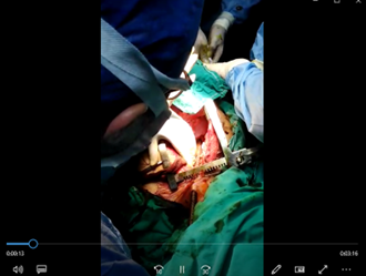
After removal of the rod, continuous bleeding started to appear from its track in the liver. Packing of the track was done using gauzes and absorbable hemostatics. Other abdominal injuries were excluded.
The right upper lung lobe tear together with the right diaphragmatic tear were repaired from the thoracic side. Closure of the thoracic cavity in layers was done after hemostasis and insertion of right pleural and pericardial drains.
Regarding the abdominal wound, only the skin was closed leaving behind a drain in Morrison’s pouch, planning for a second look after 48 hours. The patient was extubated one hour later in the ICU.
After 48 hours, the child showed no signs of active bleeding with good lab results, so abdominal reexploration was done with removal of the packs around the liver that showed complete sealing, then abdominal wound was closed in layers after drain insertion in Morrison’s pouch.
One day later, the child started enteral feeding then drains were removed and he was discharged home after 6 days of hospital stay.
Comment:
Impalement injuries in general are very rare in civilian practice [5,6]. Almost all the patients who suffer from these injuries die before arrival to the hospital [4–6]. Most likely causes are accidental falls, road traffic accidents with a protruding object or patients ejected from automobiles falling onto protruding object5. Because of the diversity in impalement mechanisms, thoracic impalement injury is always an individualized complex state in a polytraumatized patient who needs a multidisciplinary team that can manage the trauma in harmony to save his life [6]. Patient’s vital signs indicate the extent of the injury. Altough, time shouldn’t be wasted in unnecessary investigations because there might be a concealed injury that is controlled by the object inplace[7]. CT-scan with contrast is the best to inspect visceral and vascular injuries. Efforts to remove such an object should always be taken in the operating room where everything is prepared to control any injuries [6].
Impalement injuries aren’t common in the civilian practice. Once happened, the patient should be transferred to a tertiary care center where a multidisciplinary team can deal with such injuries. Unnecessary investigations should be avoided to save time which is a golden factor in such cases. A proper incision to safely trace and secure the course of the impaled object during its extraction is mandatory.
Dear Editorial Team, Clinical Medical Reviews and Reports. My experience with the journal was highly positive. The peer-review process was rigorous, constructive, and completed in a timely manner. The reviewers provided valuable comments that helped improve the quality and clarity of our manuscript. The editorial office was professional, responsive, and supportive throughout all stages of the publication process. Communication was clear and efficient, and any questions were addressed promptly. Overall, I found the journal to maintain high scientific standards and an excellent publication workflow. I would be pleased to consider submitting future work to this journal. Best wishes from, Elena Popa.

It was my pleasure to submit my testimonial concerning the Reviewer Board of our Scientific Journal “Brain and Neurological Disorders”. The Reviewers focused on some modifications and their contribution was helpful. The ladies of our Editorial Office were also supported my efforts. It was my honor to have such a co-operation and I am looking forward for more collaboration.

Dear Grace Pierce, Editorial Coordinator of Journal of Clinical Research and Reports, Thank you for the speedy and efficient peer review process. I appreciate the fact that your peer reviewers do not take months to respond like with some other journals. I would also like to thank the editorial office for responding quickly to my questions. It is an excellent journal. I plan to submit more manuscripts in the future. Best wishes from, Robert W. McGee

Dear Grace Pierce, Editorial Coordinator of Journal of Clinical Research and Reports, Working with you and your team on our recent publication in JCRR has been a truly wonderful and enjoyable experience. The responses were prompt, and the reviewers were patient, constructive, and highly professional. One reviewer in particular gave me the feeling that a professor was carefully reading and commenting on my coursework, which was deeply touching. The entire process was straightforward and hassle‑free, with no tedious online forms to complete. I highly recommend this journal. Best wishes from, DR Aibing Rao, Head of R&D

I Appreciate the Opportunity to Share my Experience with the Journal of Clinical Research and Reports. The peer review process was timely and constructive, and the feedback provided helped improve the quality of our manuscript. The editorial office was professional, responsive, and supportive throughout the process, ensuring smooth communication and efficient handling of the submission. Overall, it was a positive experience collaborating with your team.

Dear Mercy Grace, Editorial Coordinator of Obstetrics Gynecology and Reproductive Sciences, We would like to express our gratitude for your help at all stages of publishing and editing the article. The editors of the magazine answer all the necessary questions and help at every stage. We will definitely continue to cooperate and publish other works in the Obstetrics Gynecology and Reproductive Sciences! Best wishes from, Alla Konstantinovna Politova,
