AUCTORES
Globalize your Research

Research Article | DOI: https://doi.org/10.31579/2641-5194/064
1 Specialist of Internal Medicine, MD, Turkey.
2 Manager of Writing and Statistics, Turkey.
3 Ministry of Health of Turkey, MD, Turkey.
4 Specialist of Emergency Medicine, MD, Turkey.
5 Middle-East Academy for Medicine of Aging, MD, Lebanon.
6 Medi-WORLD International, Australia.
*Corresponding Author: Mehmet Rami Helvaci, Specialist of Internal Medicine, MD, Turkey.
Citation: Mehmet R. Helvaci, Kubra Piral, Kubra Seckin, Esin E. Tanaydin, Ayse D.Karabacak, Mehpare Camlibel, et al, (2023), Fasting Plasma Glucose May Behave as a Positive in Mild but as a Negative Acute Phase Reactant in Moderate and Severe Inflammatory Disorders, J. Gastroenterology Pancreatology and Hepatobilary Disorders, 7(2); DOI:10.31579/2641-5194/064
Copyright: 2023, Mehmet Rami Helvaci. This is an open access article distributed under the Creative Commons Attribution License, which permits unrestricted use, distribution, and reproduction in any medium, provided the original work is properly cited.
Received: 31 May 2023 | Accepted: 08 June 2023 | Published: 16 June 2023
Keywords: fasting plasma glucose; diabetes mellitus; irritable bowel syndrome; smoking; digital clubbing; sickle cell diseases; atherosclerosis
Background: There may be significant relationships between fasting plasma glucose (FPG) and severity of inflammations.
Method: All cases with the digital clubbing were included.
Results: The study included 104 cases with clubbing detected among 2.428 cases (1.044 males). So clubbing was higher in males (8.1% versus 1.3%, p<0.001). Mean age of clubbing cases was 49.2 years, and there was a male predominance (81.7%), again. Parallel to the male predominance, there were higher prevalences of smoking (69.2% versus 41.6%, p<0.001), chronic obstructive pulmonary disease (COPD) (27.8% versus 10.8%, p<0.001), and coronary heart disease (CHD) and/or peripheric artery disease (PAD) (7.6% versus 0.0%, p<0.01) in the clubbing cases. Whereas the body weight, body mass index (BMI), and FPG were lower in the clubbing cases but the differences were nonsignificant probably due to the small sample size. But diabetes mellitus (DM) (12.5% versus 21.6%, p<0.05) and systolic blood pressure (BP) (127.6 versus 136.9 mmHg, p= 0.011) were lower in the clubbing cases, significantly.
Conclusion: There are significant relationships between smoking, digital clubbing, COPD, CHD, and PAD probably due to strong atherosclerotic effects of smoking. Similarly, the body weight, BMI, FPG, systolic BP, and DM are inversely related with the clubbing probably due to the severe inflammatory effects of smoking on the vascular endothelium, again. FPG may behave as a positive acute phase reactant (APR) in mild inflammatory disorders such as irritable bowel syndrome but as a negative APR in moderate and severe inflammatory disorders such as smoking, digital clubbing, and sickle cell diseases.
Digital changes may help to identify some systemic disorders in human body. Digital clubbing is a deformity of the fingers and fingernails that is known for a long time. It is characterized by bulbous enlargement of the distal phalanges due to the increase in soft tissue. Digital clubbing develops in the following steps; fluctuation and softening of the nailbed, loss of normal angle between the nailbed and fold which is lower than 165°, increased convexity of the nail fold, thickening of the whole distal finger, and shiny aspect and striation of the nail and skin [1]. Schamroth’s window test is a popular test for the diagnosis of digital clubbing [2]. When the distal phalanges of corresponding fingers of opposite hands are directly opposed, a small diamond-shaped ‘window’ is apparent between the nailbeds, normally. If this window is obliterated, the test is positive and digital clubbing is present. Although many disorders may be associated with the clubbing, the reports are mostly anecdotal, and prospective studies of patients with the clubbing have not been performed, yet. The clubbing may be associated with pulmonary, cardiac, and hepatic diseases that are featuring with chronic tissue hypoxia (tuberculosis, bronchiectasis), hypothyroidism, gastrointestinal and hepatobiliary disorders (malabsorption, Crohn’s disease, ulcerative colitis, cirrhosis), thymoma, thalassemia, and human immunodeficiency virus infection [3-7]. But there was not any underlying disorder in 60% of cases [8]. Additionally, the exact prevalence of digital clubbing in the population is not known. The above study detected digital clubbing just in 0.9% of all patients admitted to the Department of Internal Medicine, and 66.6% of the clubbing cases were male [8]. Probably due to the higher prevalence of smoking in males [9], the great gender differences were observed in the clubbing. We tried to understand whether or not there are some relationships between fasting plasma glucose (FPG) and severity of inflammations in human body.
The study was performed in the Internal Medicine Clinic of the Mustafa Kemal University between March 2007 and May 2011 on all patients applying for any complaint. Their medical histories including smoking, claudication, angina pectoris, and already used medications were learnt, and a routine check-up procedure including FPG, total cholesterol, high density lipoproteins (HDL), triglycerides, an electrocardiography, and a Doppler echocardiogram just in suspected cases was performed. Digital clubbing is diagnosed by determining ratio of the distal phalangeal diameter to the interphalangeal diameter which is required to be greater than 1.0, and with the presence of the Schamroth sign [2, 8]. Current daily smokers at least for the last six months and cases with a history of five pack-years were accepted as smokers. Body mass index (BMI) of each case was calculated by the measurements of the Same Physician instead of verbal expressions (10). Office blood pressure (BP) was checked after a five-minute of rest in seated position with the mercury sphygmomanometer (ERKA, Germany). Cases with an overnight FPG level of 126 mg/dL or higher on two occasions or already using antidiabetic medications were defined as diabetics [10]. An oral glucose tolerance test with 75-gram glucose was performed in cases with a FPG level between 100 and 125 mg/dL, and diagnosis of cases with a two-hour plasma glucose level of 200 mg/dL or greater was diabetes mellitus (DM) [10]. An exercise electrocardiogram was performed just in cases with an abnormal electrocardiogram and/or angina pectoris. Coronary angiography was taken just for exercise electrocardiogram positive cases. So coronary heart disease (CHD) was diagnosed either angiographically or with the Doppler echocardiographic findings as the movement disorders of the cardiac walls. A colored Doppler ultrasonograpy of arterial system of the lower extremities were obtained just in cases with a history of claudication for the diagnosis of peripheric artery disease (PAD). Chronic obstructive pulmonary disease (COPD) was diagnosed by means of spirometric measurements. The criterion for diagnosis of COPD is post-bronchodilator forced expiratory volume in one second/forced vital capacity of less than 70% [11]. Eventually, all cases with the clubbing were collected into the first, and age- and sex-matched control cases were collected into the second groups, and compared in between. Mann-Whitney U test, Independent-Samples t test, and comparison of proportions were used as the methods of statistical analyses.
The study included 104 patients (85 males) with the digital clubbing and 120 control cases. The clubbing cases were detected among 2.428 cases (1.044 males), totally. So clubbing was higher in males, significantly (8.1% versus 1.3%, p<0 xss=removed>0.05). On the other hand, low density lipoproteins (LDL) (130.0 versus 126.9 mg/dL, p>0.05) and triglycerides (152.5 versus 143.4 mg/dL, p>0.05) were higher in the digital clubbing cases but the differences were nonsignificant probably due to the small sample size of the study, again. There were seven cases with CHD and one case with PAD in the clubbing group, whereas no case could be detected in the control group (Table 1).
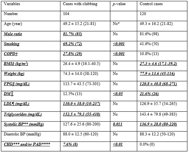
Table 1: Characteristics features of the study cases.
*Nonsignificant (p>0.05) †Chronic obstructive pulmonary disease ‡Body mass index §Fasting plasma glucose ║Diabetes mellitus ¶Low density lipoproteins **Blood pressure ***coronary heart disease ****Peripheric artery disease
Recurrent upper abdominal discomfort may be the cause of nearly half of applications to the Internal Medicine Clinics (12), and irritable bowel syndrome (IBS) and chronic gastritis may be the most commonly diagnosed disorders in such cases. Flatulence, periods of diarrhea and constipation, repeated toilet visits due to urgent evacuation or early filling sensation, excessive straining, feeling of incomplete evacuation, frequency, urgency, reduced feeling of well-being, and eventually disturbed social life are often reported by the patients with IBS. Although many patients relate onset of symptoms to intake of food, and often incriminate specific food items, a meaningful dietary role is doubtful in the IBS. According to the literature, nearly 20% of general population have IBS, and it is more common in females with unknown causes, yet [13]. Psychological factors seem to precede onset and exacerbation of gut symptoms, and many potentially psychiatric disorders including anxiety, depression, sleep disorders, illness fear, cancer fear, or death fear usually coexist with the IBS [14]. For example, thresholds for sensations of initial filling, evacuation, urgent evacuation, and utmost tolerance recorded via a rectal balloon significantly decreased by focusing the examiners' attention on gastrointestinal stimuli by reading pictures of gastrointestinal malignancies in patients with the IBS [15]. In other words, although IBS is described as a physical disorder according to Rome II guidelines, psychological factors may be crucial for triggering of these physical changes in the body. Eventually, IBS may even terminate with chronic gastritis, urolithiasis, and hemorrhoids [16-18]. Similarly, some authors studied the role of inflammation in IBS via colonic biopsies in 77 patients [19]. Although 38 patients had normal histology, 31 patients demonstrated microscopic inflammation, and eight patients fulfilled criteria for lymphocytic colitis. However, immunohistology revealed increased intraepithelial lymphocytes as well as increased CD3 and CD25 positive cells in lamina propria of the group with “normal” histology. These features were more evident in the microscopic inflammation group who additionally revealed increased neutrophils, mast cells, and natural killer cells. All of these immunopathological abnormalities were the most evident in the lymphocytic colitis group who also demonstrated HLA-DR staining in the crypts and increased CD8 positive cells in the lamina propria [19]. A direct link between the immunologic activation and IBS symptoms was shown by some other authors, too [20]. They demonstrated not only an increased mast cell degranulation in the colon but also a direct correlation between proximity of mast cells to neuronal elements and severity of pain in the IBS [20]. In addition to above findings, there are some evidences for extension of the inflammatory process behind the mucosa. Some authors addressed this issue in ten patients with severe IBS by examining full-thickness jejunal biopsies obtained via laparoscopy [21]. They detected a low-grade infiltration of lymphocytes in myenteric plexus of nine patients, four of whom had an associated increase in intraepithelial lymphocytes and six demonstrated evidence of neuronal degeneration [21]. Nine patients had hypertrophy of longitudinal muscles, and seven had abnormalities in number and size of interstitial cells of Cajal [21]. The finding of intraepithelial lymphocytosis was also consistent with some other reports in the colon [19] and duodenum [22]. So, IBS may have more complex mechanisms affecting various systems of the body by means of a low-grade inflammatory process [23]. Beside that mean values of FPG (111.9 versus 105.4 mg/dL, p= 0.002) and plasma triglycerides (167.0 versus 147.3 mg/dL, p= 0.013) were higher in the IBS cases (24). Because plasma triglycerides are well-known acute phase reactants (APRs), the additionally increased FPG in the IBS cases may show the fact that FPG may behave as a positive APR in the IBS case (24).
Sickle cell diseases (SCDs) are chronic inflammatory process on vascular endothelium, initiated at birth and terminated with an accelerated
atherosclerosis induced end-organ failures in early years of life [25, 26]. Hemoglobin S causes loss of elastic and biconcave disc shaped structures of red blood cells (RBCs). Probably loss of elasticity is the main problem instead of the shape since sickling is rare in peripheric blood samples of the patients with associated thalassemia minors, and human survival is not affected in hereditary spherocytosis or elliptocytosis. Loss of elasticity is present during whole lifespan, but exaggerated with inflammation, infection, depression, and various stresses of the body. The hardened RBCs induced chronic endothelial damage, inflammation, and fibrosis terminate with disseminated tissue hypoxia all over the body [27]. As a difference from other causes of chronic endothelial damage, the SCDs may keep vascular endothelium particularly at the capillary level [28, 29], since the capillary system is the main distributor of the hardened RBCs into the tissues. The hardened RBCs induced chronic endothelial damage builds up an advanced atherosclerosis in early years of life. Vascular narrowings and occlusions induced tissue ischemia and infarctions are the final consequences, so the mean life expectancy is decreased by 25 to 30 years in both genders in the SCDs [26]. Due to the severity of inflammation in the SCDs, the body weight, BMI, FPG, LDL, HDL, systolic and diastolic BPs, and hematocrit values decreased as some negative APRs in the body, significantly [30].
The monolayer of endothelial cells that forms the inner lining of arteries, veins, capillaries, and lymphatics is called as the endothelium. Probably, the whole endothelium all over the body may act as a private organ that may be the largest organ of the body. It may contract vasculature of the peripheral organs while relaxing the internal ones during cold, anxiety, and depression-like stresses. Because we measure the systolic and diastolic BPs of the arms and legs, they may not show the actual BPs of the brain, heart, lung, liver, and kidney-like internal organs. The endothelium may be the main organ in the control of blood fluidity, platelets aggregation, and vascular tone in the body. It may control vascular tone and blood flow by releasing nitric oxide, reactive oxygen species, and metabolites of arachidonic acid into the circulation. It may also be important for synthesizing of vasoactive hormones such as angiotensin II. An endothelial dysfunction-induced accelerated atherosclerosis all over the body may be the main cause of end-organ insufficiencies, aging, and death. Such a dysfunction may also be important in the development of cancers by preventing clearance of malignant cells by the natural killers in terminal points of the circulation. Similarly, physical inactivity, animal-rich diet, excess weight, higher BP and glucose levels, chronic inflammations, prolonged infections, cancers, smoking, and alcohol may be accelerating factors of the chronic endothelial inflammation and dysfunction terminating with the accelerated atherosclerosis-induced end-organ insufficiencies (31). The much higher BPs of the afferent vasculature may be the major accelerating factor by inducing recurrent injuries on the vascular endothelium. Probably, whole afferent vasculature including capillaries are mainly involved in the process. Thus the term of venosclerosis is not as famous as atherosclerosis in the literature. Due to the chronic endothelial damage, inflammation, edema, fibrosis, and dysfunction, vascular walls thicken, their lumens narrow, and they lose their elastic natures, those eventually reduce blood flow to the terminal organs, and increase systolic and decrease diastolic BPs further. Some of the irreversible consequences of the systemic inflammatory process are obesity, hypertension (HT), DM, cirrhosis, PAD, COPD, CHD, chronic renal disease (CRD), mesenteric ischemia, osteoporosis, stroke, dementia, aging, and death (32). Although early withdrawal of the accelerating factors may delay terminal consequences, endothelial changes cannot be reversed, completely after development of the irreversible end-points due to their fibrotic natures. The accelerating factors and irreversible end-points are researched under the titles of metabolic syndrome, aging syndrome, and accelerated endothelial damage syndrome, extensively [33, 34].
Obesity may be one of the irreversible end-points of the metabolic syndrome. Although some transient successes can be achieved, nonpharmaceutical approaches provide limited benefit to reverse the obesity, permanently. Due to the excess weight-induced chronic low-grade inflammation on the vascular endothelium, the risk of death from all causes including cardiovascular diseases and cancers increases parallel to the range of excess weight in all age groups [35]. The chronic low-grade inflammation may even cause genetic changes of the endothelial cells, and the systemic atherosclerosis may prevent clearance of malignant cells, effectively. Similarly, the effects of excess weight on the BP were shown in the literature, extensively (36). For example, prevalences of sustained normotension (NT) were higher in the underweight than the normal weight (80.3% versus 64.0%, p<0>
Smoking may be the second common cause of disseminated vasculitis in human body. It may cause a low-grade systemic inflammation on vascular endothelium terminating with an accelerated atherosclerosis-induced end-organ insufficiencies all over the body [42]. Its atherosclerotic effect is the most obvious in Buerger’s disease. Buerger’s disease is an obliterative vasculitis characterized by inflammatory changes in the small and medium-sized arteries and veins, and it has never been reported in the absence of smoking. Plasma triglycerides, LDL, erythrocyte sedimentation rate (ESR), and C-reactive protein (CRP) may be positive whereas HDL and FPG may be negative APRs indicating such inflammatory effects of smoking in the body [43]. Parallel to the systemic inflammatory and atherosclerotic effects of smoking, smoking in human being and nicotine administration in animals were associated with the lower values of BMI in some studies [44]. Some evidences revealed an increased energy expenditure during smoking both on the rest and light physical activity [45]. Nicotine supplied by patch after smoking cessation decreased caloric intake in a dose-related manner [46]. According to an animal study, nicotine may lengthen intermeal time, and decrease amount of meal eaten [47]. Smoking may be associated with a postcessation weight gain, but the risk is the highest during the first year, and decreases with the following years [48]. As the opposite findings to the above studies, the mean weight and BMI were similar both in the smokers and non-smokers in the other study [43]. Similarly, prevalences of smoking were similar in the normal weight (35.9%), overweight (32.9%), and obesity groups (33.7%, p>0.05 between all) in another study [49]. On the other hand, although the CHD was detected with similar prevalences in both genders, prevalences of smoking and COPD were higher in males against the higher BMI, LDL, triglycerides, WCH, HT, and DM in females [50]. Beside that the prevalence of myocardial infarctions is increased three-fold in men and six-fold in women who smoked at least 20 cigarettes per day [51]. In another word, smoking may be more dangerous for women about the atherosclerotic end-points probably due to the higher BMI and its consequences in them. Several toxic substances found in the cigarette smoke get into the circulation, and cause the vascular endothelial inflammation in various organ systems of the body. For example, smoking is usually reported together with depression, IBS, chronic gastritis, hemorrhoids, and urolithiasis in the literature (16, 52). There may be several underlying mechanisms to explain these associations in the smokers [52]. First of all, smoking may have some additional antidepressant properties with several side effects. Secondly, smoking-induced vascular endothelial inflammation may disturb epithelial functions for absorption and excretion in the gastrointestinal and genitourinary tracts. These functional problems may terminate with urolithiasis and components of the IBS including loose stool, diarrhea, and constipation. Thirdly, diarrheal losses-induced urinary changes may even cause urolithiasis [16, 17]. Fourthly, smoking-induced sympathetic nervous system activation may cause motility problems in the gastrointestinal and genitourinary tracts terminating with the IBS and urolithiasis. Eventually, immunosuppression secondary to smoking-induced vascular endothelial inflammation may even terminate with the gastrointestinal and genitourinary tract infections causing loose stool, diarrhea, and urolithiasis, because some types of bacteria can provoke urinary supersaturation, and modify the environment to form crystal deposits in the urine. Actually, 10% of urinary stones are struvite stones which are built by magnesium ammonium phosphate produced during infections with the bacteria producing urease. Parallel to the results above, urolithiasis was detected in 17.9% of cases with the IBS and 11.6% of cases without in the other study (p<0>
Alcohol may be the third common cause of systemic vasculitis in human body. It is addictive to humans, and can result in alcohol use disorder (AUD), dependence, and withdrawal. Alcohol is causally associated with more than 200 different pathologies including cancers in whole body [53]. Eventually, people hospitalized with AUD have an average life expectancy of 47-53 years in men and 50-58 years in women, and die 24-28 years earlier than the others [54]. People with AUD have three-fold higher mortality in men and four-fold in women [55]. Similar to smoking, alcohol may be more dangerous for women about the atherosclerotic end-points probably due to their lower body mass induced lower capacity to metabolize alcohol and higher body fat. A very substantial part of the Danish excess mortality and lower life expectancy compared to Sweden can be attributed to higher mortality related with alcohol and smoking [54]. It may even cause unconsciousness and sudden death if taken in high amounts. Hepatic alcohol dehydrogenase is the main enzyme to metabolize alcohol that requires the cofactor, nicotinamide adenine dinucleotide (NAD). Normally, NAD is used to metabolize fats in the liver but alcohol competes with these fats for the use of NAD. Eventually, prolonged exposure of alcohol causes fatty liver. Ethanol is the only alcohol that is found in alcoholic beverages. Ethanol crosses biological membranes and blood-brain barrier by means of the passive diffusion, easily. Alcohol works particularly by increasing effects of the gamma aminobutyric acid that is the main inhibitory neurotransmitter of the brain. Alcohol causes happiness and euphoria, decreased anxiety, increased sociability, sedation, generalized depression of central nervous system, and impairment of cognitive, memory, motor, and sensory functions. It may even cause fetal disorders in pregnancy since ethanol is classified as a teratogen. Regular alcohol consumption leads to cell death in the liver, scarring, cirrhosis, and hepatocellular carcinoma. Heavy consumption may even terminate with permanent brain damage. Alcohol is the major contributing factor of elevated triglycerides which are the sensitive APRs in the plasma (38). Although regular alcohol consumers were excluded, plasma triglycerides were higher in the smokers (163.1 versus 151.3 mg/dL, p<0>
The acute phase response occurs in case of infection, infarction, cancer, trauma, depression, and burn-like inflammatory conditions of the body. Certain mediators known as APRs are increased or decreased during the response [57, 58]. These markers are commonly used in the clinical practice as the indicators of acute and chronic inflammations in the body. The terms of acute phase proteins and APRs are usually used synonymously, although some APRs are polypeptides rather than proteins. Positive and negative APRs are those whose concentrations increase or decrease during the acute phase response, respectively. The response is predominantly mediated by the pro-inflammatory cytokines including TNF, interleukin-1, and interleukin-6 secreted by neutrophils and macrophages into the circulation. The liver and other organs respond to the cytokines by producing many positive APRs. ESR, CRP, fibrinogen, ferritin, procalcitonin, hepcidin, haptoglobin, ceruloplasmin, complement proteins, and serum amyloid A are some of the well-known positive APRs. CRP is a useful indicator of the acute phase response, clinically. It is responsible for activation of the complement pathway. CRP reaches up to the maximum concentration within two days, and decreases with the resolution of the inflammation with a half-life of 6-8 hours, rapidly. It correlates with ESR, but not simultaneously since ESR is largely dependent upon elevation of fibrinogen with a half-life of one week, approximately. Thus, ESR remains higher for a longer period of time despite the removal of the inflammatory stimulus. Similarly, white blood cells and platelet counts may also behave as some other positive APRs in the body [59]. On the other hand, productions of the negative APRs are suppressed, simultaneously. Albumin, transferrin, retinol-binding protein, antithrombin, transcortin, alpha-fetoprotein, and hemoglobin are some of the well-known negative APRs in the body. Suppressions of such negative APRs are also used as the indicators of the acute phase response in the body. Suppressions of such negative APRs may actually be secondary to the protection of amino acids and polypeptides required for the production of positive APRs, sufficiently. As also observed in the smokers in the above study (56), production of HDL may also be suppressed in the liver during the acute phase response [60]. Similarly, triglycerides, DM, and CHD were all higher in patients with plasma HDL values of lower than 40 mg/dL, significantly [60]. So, HDL may actually behave as negative whereas triglycerides positive APRs in the plasma. Similarly, the highest CHD of the group with HDL values of lower than 40 mg/dL can also be explained by the same hypothesis in the other study (38). Additionally, plasma triglycerides increased whereas HDL decreased during infections [61]. On the other hand, a 10 mg/dL increase of plasma LDL values was associated with a 3% lower risk of hemorrhagic stroke [62]. Similarly, the highest prevalences of HT and DM parallel to the elevated values of LDL and HDL, and the highest prevalences of COPD, CHD, and CRD in contrast to the lowest values of LDL and HDL may show initially positive but eventually negative behaviors of LDL and HDL as the APRs [63]. Probably, HDL turn to the negative direction much earlier than LDL in the plasma. Interestingly, the most desired values were between 80 and 100 mg/dL for LDL, between 40 and 46 mg/dL for HDL, and lower than 60 mg/dL for triglycerides in the plasma [38]. Parallel to ESR and CRP, plasma triglycerides and LDL may behave as positive whereas FPG and HDL negative APRs in smokers [56]. In another word, lower HDL values should alert clinicians for researching of any acute phase response in the body [64, 65].
Cholesterol, triglycerides, and phospholipids are the major lipids of the body. They do not circulate in the plasma, freely instead they are bound to proteins, and transported as lipoproteins. There are five mjor classes of lipoproteins in the plasma. Chylomicrons carry exogenous triglycerides to the liver via the thorasic duct. Very low-density lipoproteins (VLDL) are produced in the liver, and carry endogenous triglycerides to the organs. VLDL are converted into the intermediate density lipoproteins (IDL) by removal of 90% of triglycerides by lipases in the capillaries of adipocytes and muscle tissues. Then the IDL are degraded into LDL by removal of more triglycerides. So VLDL are the main source of LDL in the plasma, and LDL deliver cholesterol from the liver to organs. Although the liver removes majority of LDL from the circulation, a small amount is uptaken by scavenger receptors of the macrophages migrating into the arterial walls, and become the foam cells of atherosclerotic plaques. HDL remove fats and cholesterol from cells including the arterial wall atheroma, and carry the cholesterol back to the adrenals, ovaries, and testes-like steroidogenic organs and liver for excretion, re-utilization, or disposal. All of the carrier lipoproteins are under dynamic control, and are readily affected by diet, drugs, inflammations, infections, cancers, trauma, smoking, alcohol, and excess weight. Thus, lipid analysis should be performed during a steady state. For example, the metabolic syndrome alone is a low-grade inflammatory process, and it may even cause abnormal lipoproteins levels in the plasma. HDL may normally show various anti-oxidative, anti-inflammatory, and anti-atherogenic properties including reverse cholesterol transport [66]. However, HDL may become ‘dysfunctional’ in pathologic conditions which means that relative compositions of lipids and proteins, as well as the enzymatic activities of HDL are altered (66). For example, properties of HDL are compromised in patients with DM by means of the oxidative modification, glycation, and/or transformation of HDL proteomes into the proinflammatory proteins. Additionally, the drugs increasing HDL values such as niacin, fibrates, and cholesteryl ester transfer protein inhibitors cannot reduce all-cause mortality, CHD mortality, myocardial infarction, and stroke [67]. In other words, HDL may just be some indicators instead of being the main actors of the health. Similarly, BMI, DM, and CHD were the lowest between the HDL values of 40 and 46 mg/dL, and the prevalence of DM was only 3.1
Clearly Auctoresonline and particularly Psychology and Mental Health Care Journal is dedicated to improving health care services for individuals and populations. The editorial boards' ability to efficiently recognize and share the global importance of health literacy with a variety of stakeholders. Auctoresonline publishing platform can be used to facilitate of optimal client-based services and should be added to health care professionals' repertoire of evidence-based health care resources.

Journal of Clinical Cardiology and Cardiovascular Intervention The submission and review process was adequate. However I think that the publication total value should have been enlightened in early fases. Thank you for all.

Journal of Women Health Care and Issues By the present mail, I want to say thank to you and tour colleagues for facilitating my published article. Specially thank you for the peer review process, support from the editorial office. I appreciate positively the quality of your journal.

Journal of Clinical Research and Reports I would be very delighted to submit my testimonial regarding the reviewer board and the editorial office. The reviewer board were accurate and helpful regarding any modifications for my manuscript. And the editorial office were very helpful and supportive in contacting and monitoring with any update and offering help. It was my pleasure to contribute with your promising Journal and I am looking forward for more collaboration.

We would like to thank the Journal of Thoracic Disease and Cardiothoracic Surgery because of the services they provided us for our articles. The peer-review process was done in a very excellent time manner, and the opinions of the reviewers helped us to improve our manuscript further. The editorial office had an outstanding correspondence with us and guided us in many ways. During a hard time of the pandemic that is affecting every one of us tremendously, the editorial office helped us make everything easier for publishing scientific work. Hope for a more scientific relationship with your Journal.

The peer-review process which consisted high quality queries on the paper. I did answer six reviewers’ questions and comments before the paper was accepted. The support from the editorial office is excellent.

Journal of Neuroscience and Neurological Surgery. I had the experience of publishing a research article recently. The whole process was simple from submission to publication. The reviewers made specific and valuable recommendations and corrections that improved the quality of my publication. I strongly recommend this Journal.

Dr. Katarzyna Byczkowska My testimonial covering: "The peer review process is quick and effective. The support from the editorial office is very professional and friendly. Quality of the Clinical Cardiology and Cardiovascular Interventions is scientific and publishes ground-breaking research on cardiology that is useful for other professionals in the field.

Thank you most sincerely, with regard to the support you have given in relation to the reviewing process and the processing of my article entitled "Large Cell Neuroendocrine Carcinoma of The Prostate Gland: A Review and Update" for publication in your esteemed Journal, Journal of Cancer Research and Cellular Therapeutics". The editorial team has been very supportive.

Testimony of Journal of Clinical Otorhinolaryngology: work with your Reviews has been a educational and constructive experience. The editorial office were very helpful and supportive. It was a pleasure to contribute to your Journal.

Dr. Bernard Terkimbi Utoo, I am happy to publish my scientific work in Journal of Women Health Care and Issues (JWHCI). The manuscript submission was seamless and peer review process was top notch. I was amazed that 4 reviewers worked on the manuscript which made it a highly technical, standard and excellent quality paper. I appreciate the format and consideration for the APC as well as the speed of publication. It is my pleasure to continue with this scientific relationship with the esteem JWHCI.

This is an acknowledgment for peer reviewers, editorial board of Journal of Clinical Research and Reports. They show a lot of consideration for us as publishers for our research article “Evaluation of the different factors associated with side effects of COVID-19 vaccination on medical students, Mutah university, Al-Karak, Jordan”, in a very professional and easy way. This journal is one of outstanding medical journal.

Dear Hao Jiang, to Journal of Nutrition and Food Processing We greatly appreciate the efficient, professional and rapid processing of our paper by your team. If there is anything else we should do, please do not hesitate to let us know. On behalf of my co-authors, we would like to express our great appreciation to editor and reviewers.

As an author who has recently published in the journal "Brain and Neurological Disorders". I am delighted to provide a testimonial on the peer review process, editorial office support, and the overall quality of the journal. The peer review process at Brain and Neurological Disorders is rigorous and meticulous, ensuring that only high-quality, evidence-based research is published. The reviewers are experts in their fields, and their comments and suggestions were constructive and helped improve the quality of my manuscript. The review process was timely and efficient, with clear communication from the editorial office at each stage. The support from the editorial office was exceptional throughout the entire process. The editorial staff was responsive, professional, and always willing to help. They provided valuable guidance on formatting, structure, and ethical considerations, making the submission process seamless. Moreover, they kept me informed about the status of my manuscript and provided timely updates, which made the process less stressful. The journal Brain and Neurological Disorders is of the highest quality, with a strong focus on publishing cutting-edge research in the field of neurology. The articles published in this journal are well-researched, rigorously peer-reviewed, and written by experts in the field. The journal maintains high standards, ensuring that readers are provided with the most up-to-date and reliable information on brain and neurological disorders. In conclusion, I had a wonderful experience publishing in Brain and Neurological Disorders. The peer review process was thorough, the editorial office provided exceptional support, and the journal's quality is second to none. I would highly recommend this journal to any researcher working in the field of neurology and brain disorders.
Dear Agrippa Hilda, Journal of Neuroscience and Neurological Surgery, Editorial Coordinator, I trust this message finds you well. I want to extend my appreciation for considering my article for publication in your esteemed journal. I am pleased to provide a testimonial regarding the peer review process and the support received from your editorial office. The peer review process for my paper was carried out in a highly professional and thorough manner. The feedback and comments provided by the authors were constructive and very useful in improving the quality of the manuscript. This rigorous assessment process undoubtedly contributes to the high standards maintained by your journal.

International Journal of Clinical Case Reports and Reviews. I strongly recommend to consider submitting your work to this high-quality journal. The support and availability of the Editorial staff is outstanding and the review process was both efficient and rigorous.

Thank you very much for publishing my Research Article titled “Comparing Treatment Outcome Of Allergic Rhinitis Patients After Using Fluticasone Nasal Spray And Nasal Douching" in the Journal of Clinical Otorhinolaryngology. As Medical Professionals we are immensely benefited from study of various informative Articles and Papers published in this high quality Journal. I look forward to enriching my knowledge by regular study of the Journal and contribute my future work in the field of ENT through the Journal for use by the medical fraternity. The support from the Editorial office was excellent and very prompt. I also welcome the comments received from the readers of my Research Article.

Dear Erica Kelsey, Editorial Coordinator of Cancer Research and Cellular Therapeutics Our team is very satisfied with the processing of our paper by your journal. That was fast, efficient, rigorous, but without unnecessary complications. We appreciated the very short time between the submission of the paper and its publication on line on your site.

I am very glad to say that the peer review process is very successful and fast and support from the Editorial Office. Therefore, I would like to continue our scientific relationship for a long time. And I especially thank you for your kindly attention towards my article. Have a good day!

"We recently published an article entitled “Influence of beta-Cyclodextrins upon the Degradation of Carbofuran Derivatives under Alkaline Conditions" in the Journal of “Pesticides and Biofertilizers” to show that the cyclodextrins protect the carbamates increasing their half-life time in the presence of basic conditions This will be very helpful to understand carbofuran behaviour in the analytical, agro-environmental and food areas. We greatly appreciated the interaction with the editor and the editorial team; we were particularly well accompanied during the course of the revision process, since all various steps towards publication were short and without delay".

I would like to express my gratitude towards you process of article review and submission. I found this to be very fair and expedient. Your follow up has been excellent. I have many publications in national and international journal and your process has been one of the best so far. Keep up the great work.

We are grateful for this opportunity to provide a glowing recommendation to the Journal of Psychiatry and Psychotherapy. We found that the editorial team were very supportive, helpful, kept us abreast of timelines and over all very professional in nature. The peer review process was rigorous, efficient and constructive that really enhanced our article submission. The experience with this journal remains one of our best ever and we look forward to providing future submissions in the near future.

I am very pleased to serve as EBM of the journal, I hope many years of my experience in stem cells can help the journal from one way or another. As we know, stem cells hold great potential for regenerative medicine, which are mostly used to promote the repair response of diseased, dysfunctional or injured tissue using stem cells or their derivatives. I think Stem Cell Research and Therapeutics International is a great platform to publish and share the understanding towards the biology and translational or clinical application of stem cells.

I would like to give my testimony in the support I have got by the peer review process and to support the editorial office where they were of asset to support young author like me to be encouraged to publish their work in your respected journal and globalize and share knowledge across the globe. I really give my great gratitude to your journal and the peer review including the editorial office.

I am delighted to publish our manuscript entitled "A Perspective on Cocaine Induced Stroke - Its Mechanisms and Management" in the Journal of Neuroscience and Neurological Surgery. The peer review process, support from the editorial office, and quality of the journal are excellent. The manuscripts published are of high quality and of excellent scientific value. I recommend this journal very much to colleagues.

Dr.Tania Muñoz, My experience as researcher and author of a review article in The Journal Clinical Cardiology and Interventions has been very enriching and stimulating. The editorial team is excellent, performs its work with absolute responsibility and delivery. They are proactive, dynamic and receptive to all proposals. Supporting at all times the vast universe of authors who choose them as an option for publication. The team of review specialists, members of the editorial board, are brilliant professionals, with remarkable performance in medical research and scientific methodology. Together they form a frontline team that consolidates the JCCI as a magnificent option for the publication and review of high-level medical articles and broad collective interest. I am honored to be able to share my review article and open to receive all your comments.

“The peer review process of JPMHC is quick and effective. Authors are benefited by good and professional reviewers with huge experience in the field of psychology and mental health. The support from the editorial office is very professional. People to contact to are friendly and happy to help and assist any query authors might have. Quality of the Journal is scientific and publishes ground-breaking research on mental health that is useful for other professionals in the field”.

Dear editorial department: On behalf of our team, I hereby certify the reliability and superiority of the International Journal of Clinical Case Reports and Reviews in the peer review process, editorial support, and journal quality. Firstly, the peer review process of the International Journal of Clinical Case Reports and Reviews is rigorous, fair, transparent, fast, and of high quality. The editorial department invites experts from relevant fields as anonymous reviewers to review all submitted manuscripts. These experts have rich academic backgrounds and experience, and can accurately evaluate the academic quality, originality, and suitability of manuscripts. The editorial department is committed to ensuring the rigor of the peer review process, while also making every effort to ensure a fast review cycle to meet the needs of authors and the academic community. Secondly, the editorial team of the International Journal of Clinical Case Reports and Reviews is composed of a group of senior scholars and professionals with rich experience and professional knowledge in related fields. The editorial department is committed to assisting authors in improving their manuscripts, ensuring their academic accuracy, clarity, and completeness. Editors actively collaborate with authors, providing useful suggestions and feedback to promote the improvement and development of the manuscript. We believe that the support of the editorial department is one of the key factors in ensuring the quality of the journal. Finally, the International Journal of Clinical Case Reports and Reviews is renowned for its high- quality articles and strict academic standards. The editorial department is committed to publishing innovative and academically valuable research results to promote the development and progress of related fields. The International Journal of Clinical Case Reports and Reviews is reasonably priced and ensures excellent service and quality ratio, allowing authors to obtain high-level academic publishing opportunities in an affordable manner. I hereby solemnly declare that the International Journal of Clinical Case Reports and Reviews has a high level of credibility and superiority in terms of peer review process, editorial support, reasonable fees, and journal quality. Sincerely, Rui Tao.

Clinical Cardiology and Cardiovascular Interventions I testity the covering of the peer review process, support from the editorial office, and quality of the journal.

Clinical Cardiology and Cardiovascular Interventions, we deeply appreciate the interest shown in our work and its publication. It has been a true pleasure to collaborate with you. The peer review process, as well as the support provided by the editorial office, have been exceptional, and the quality of the journal is very high, which was a determining factor in our decision to publish with you.

The peer reviewers process is quick and effective, the supports from editorial office is excellent, the quality of journal is high. I would like to collabroate with Internatioanl journal of Clinical Case Reports and Reviews journal clinically in the future time.

Clinical Cardiology and Cardiovascular Interventions, I would like to express my sincerest gratitude for the trust placed in our team for the publication in your journal. It has been a true pleasure to collaborate with you on this project. I am pleased to inform you that both the peer review process and the attention from the editorial coordination have been excellent. Your team has worked with dedication and professionalism to ensure that your publication meets the highest standards of quality. We are confident that this collaboration will result in mutual success, and we are eager to see the fruits of this shared effort.

Dear Dr. Jessica Magne, Editorial Coordinator 0f Clinical Cardiology and Cardiovascular Interventions, I hope this message finds you well. I want to express my utmost gratitude for your excellent work and for the dedication and speed in the publication process of my article titled "Navigating Innovation: Qualitative Insights on Using Technology for Health Education in Acute Coronary Syndrome Patients." I am very satisfied with the peer review process, the support from the editorial office, and the quality of the journal. I hope we can maintain our scientific relationship in the long term.

Dear Monica Gissare, - Editorial Coordinator of Nutrition and Food Processing. ¨My testimony with you is truly professional, with a positive response regarding the follow-up of the article and its review, you took into account my qualities and the importance of the topic¨.

Dear Dr. Jessica Magne, Editorial Coordinator 0f Clinical Cardiology and Cardiovascular Interventions, The review process for the article “The Handling of Anti-aggregants and Anticoagulants in the Oncologic Heart Patient Submitted to Surgery” was extremely rigorous and detailed. From the initial submission to the final acceptance, the editorial team at the “Journal of Clinical Cardiology and Cardiovascular Interventions” demonstrated a high level of professionalism and dedication. The reviewers provided constructive and detailed feedback, which was essential for improving the quality of our work. Communication was always clear and efficient, ensuring that all our questions were promptly addressed. The quality of the “Journal of Clinical Cardiology and Cardiovascular Interventions” is undeniable. It is a peer-reviewed, open-access publication dedicated exclusively to disseminating high-quality research in the field of clinical cardiology and cardiovascular interventions. The journal's impact factor is currently under evaluation, and it is indexed in reputable databases, which further reinforces its credibility and relevance in the scientific field. I highly recommend this journal to researchers looking for a reputable platform to publish their studies.

Dear Editorial Coordinator of the Journal of Nutrition and Food Processing! "I would like to thank the Journal of Nutrition and Food Processing for including and publishing my article. The peer review process was very quick, movement and precise. The Editorial Board has done an extremely conscientious job with much help, valuable comments and advices. I find the journal very valuable from a professional point of view, thank you very much for allowing me to be part of it and I would like to participate in the future!”

Dealing with The Journal of Neurology and Neurological Surgery was very smooth and comprehensive. The office staff took time to address my needs and the response from editors and the office was prompt and fair. I certainly hope to publish with this journal again.Their professionalism is apparent and more than satisfactory. Susan Weiner

My Testimonial Covering as fellowing: Lin-Show Chin. The peer reviewers process is quick and effective, the supports from editorial office is excellent, the quality of journal is high. I would like to collabroate with Internatioanl journal of Clinical Case Reports and Reviews.

My experience publishing in Psychology and Mental Health Care was exceptional. The peer review process was rigorous and constructive, with reviewers providing valuable insights that helped enhance the quality of our work. The editorial team was highly supportive and responsive, making the submission process smooth and efficient. The journal's commitment to high standards and academic rigor makes it a respected platform for quality research. I am grateful for the opportunity to publish in such a reputable journal.

My experience publishing in International Journal of Clinical Case Reports and Reviews was exceptional. I Come forth to Provide a Testimonial Covering the Peer Review Process and the editorial office for the Professional and Impartial Evaluation of the Manuscript.

I would like to offer my testimony in the support. I have received through the peer review process and support the editorial office where they are to support young authors like me, encourage them to publish their work in your esteemed journals, and globalize and share knowledge globally. I really appreciate your journal, peer review, and editorial office.

Dear Agrippa Hilda- Editorial Coordinator of Journal of Neuroscience and Neurological Surgery, "The peer review process was very quick and of high quality, which can also be seen in the articles in the journal. The collaboration with the editorial office was very good."

I would like to express my sincere gratitude for the support and efficiency provided by the editorial office throughout the publication process of my article, “Delayed Vulvar Metastases from Rectal Carcinoma: A Case Report.” I greatly appreciate the assistance and guidance I received from your team, which made the entire process smooth and efficient. The peer review process was thorough and constructive, contributing to the overall quality of the final article. I am very grateful for the high level of professionalism and commitment shown by the editorial staff, and I look forward to maintaining a long-term collaboration with the International Journal of Clinical Case Reports and Reviews.

To Dear Erin Aust, I would like to express my heartfelt appreciation for the opportunity to have my work published in this esteemed journal. The entire publication process was smooth and well-organized, and I am extremely satisfied with the final result. The Editorial Team demonstrated the utmost professionalism, providing prompt and insightful feedback throughout the review process. Their clear communication and constructive suggestions were invaluable in enhancing my manuscript, and their meticulous attention to detail and dedication to quality are truly commendable. Additionally, the support from the Editorial Office was exceptional. From the initial submission to the final publication, I was guided through every step of the process with great care and professionalism. The team's responsiveness and assistance made the entire experience both easy and stress-free. I am also deeply impressed by the quality and reputation of the journal. It is an honor to have my research featured in such a respected publication, and I am confident that it will make a meaningful contribution to the field.

"I am grateful for the opportunity of contributing to [International Journal of Clinical Case Reports and Reviews] and for the rigorous review process that enhances the quality of research published in your esteemed journal. I sincerely appreciate the time and effort of your team who have dedicatedly helped me in improvising changes and modifying my manuscript. The insightful comments and constructive feedback provided have been invaluable in refining and strengthening my work".

I thank the ‘Journal of Clinical Research and Reports’ for accepting this article for publication. This is a rigorously peer reviewed journal which is on all major global scientific data bases. I note the review process was prompt, thorough and professionally critical. It gave us an insight into a number of important scientific/statistical issues. The review prompted us to review the relevant literature again and look at the limitations of the study. The peer reviewers were open, clear in the instructions and the editorial team was very prompt in their communication. This journal certainly publishes quality research articles. I would recommend the journal for any future publications.

Dear Jessica Magne, with gratitude for the joint work. Fast process of receiving and processing the submitted scientific materials in “Clinical Cardiology and Cardiovascular Interventions”. High level of competence of the editors with clear and correct recommendations and ideas for enriching the article.

We found the peer review process quick and positive in its input. The support from the editorial officer has been very agile, always with the intention of improving the article and taking into account our subsequent corrections.

My article, titled 'No Way Out of the Smartphone Epidemic Without Considering the Insights of Brain Research,' has been republished in the International Journal of Clinical Case Reports and Reviews. The review process was seamless and professional, with the editors being both friendly and supportive. I am deeply grateful for their efforts.

To Dear Erin Aust – Editorial Coordinator of Journal of General Medicine and Clinical Practice! I declare that I am absolutely satisfied with your work carried out with great competence in following the manuscript during the various stages from its receipt, during the revision process to the final acceptance for publication. Thank Prof. Elvira Farina

Dear Jessica, and the super professional team of the ‘Clinical Cardiology and Cardiovascular Interventions’ I am sincerely grateful to the coordinated work of the journal team for the no problem with the submission of my manuscript: “Cardiometabolic Disorders in A Pregnant Woman with Severe Preeclampsia on the Background of Morbid Obesity (Case Report).” The review process by 5 experts was fast, and the comments were professional, which made it more specific and academic, and the process of publication and presentation of the article was excellent. I recommend that my colleagues publish articles in this journal, and I am interested in further scientific cooperation. Sincerely and best wishes, Dr. Oleg Golyanovskiy.

Dear Ashley Rosa, Editorial Coordinator of the journal - Psychology and Mental Health Care. " The process of obtaining publication of my article in the Psychology and Mental Health Journal was positive in all areas. The peer review process resulted in a number of valuable comments, the editorial process was collaborative and timely, and the quality of this journal has been quickly noticed, resulting in alternative journals contacting me to publish with them." Warm regards, Susan Anne Smith, PhD. Australian Breastfeeding Association.

Dear Jessica Magne, Editorial Coordinator, Clinical Cardiology and Cardiovascular Interventions, Auctores Publishing LLC. I appreciate the journal (JCCI) editorial office support, the entire team leads were always ready to help, not only on technical front but also on thorough process. Also, I should thank dear reviewers’ attention to detail and creative approach to teach me and bring new insights by their comments. Surely, more discussions and introduction of other hemodynamic devices would provide better prevention and management of shock states. Your efforts and dedication in presenting educational materials in this journal are commendable. Best wishes from, Farahnaz Fallahian.

Dear Maria Emerson, Editorial Coordinator, International Journal of Clinical Case Reports and Reviews, Auctores Publishing LLC. I am delighted to have published our manuscript, "Acute Colonic Pseudo-Obstruction (ACPO): A rare but serious complication following caesarean section." I want to thank the editorial team, especially Maria Emerson, for their prompt review of the manuscript, quick responses to queries, and overall support. Yours sincerely Dr. Victor Olagundoye.

Dear Ashley Rosa, Editorial Coordinator, International Journal of Clinical Case Reports and Reviews. Many thanks for publishing this manuscript after I lost confidence the editors were most helpful, more than other journals Best wishes from, Susan Anne Smith, PhD. Australian Breastfeeding Association.

Dear Agrippa Hilda, Editorial Coordinator, Journal of Neuroscience and Neurological Surgery. The entire process including article submission, review, revision, and publication was extremely easy. The journal editor was prompt and helpful, and the reviewers contributed to the quality of the paper. Thank you so much! Eric Nussbaum, MD

Dr Hala Al Shaikh This is to acknowledge that the peer review process for the article ’ A Novel Gnrh1 Gene Mutation in Four Omani Male Siblings, Presentation and Management ’ sent to the International Journal of Clinical Case Reports and Reviews was quick and smooth. The editorial office was prompt with easy communication.

Dear Erin Aust, Editorial Coordinator, Journal of General Medicine and Clinical Practice. We are pleased to share our experience with the “Journal of General Medicine and Clinical Practice”, following the successful publication of our article. The peer review process was thorough and constructive, helping to improve the clarity and quality of the manuscript. We are especially thankful to Ms. Erin Aust, the Editorial Coordinator, for her prompt communication and continuous support throughout the process. Her professionalism ensured a smooth and efficient publication experience. The journal upholds high editorial standards, and we highly recommend it to fellow researchers seeking a credible platform for their work. Best wishes By, Dr. Rakhi Mishra.

Dear Jessica Magne, Editorial Coordinator, Clinical Cardiology and Cardiovascular Interventions, Auctores Publishing LLC. The peer review process of the journal of Clinical Cardiology and Cardiovascular Interventions was excellent and fast, as was the support of the editorial office and the quality of the journal. Kind regards Walter F. Riesen Prof. Dr. Dr. h.c. Walter F. Riesen.

Dear Ashley Rosa, Editorial Coordinator, International Journal of Clinical Case Reports and Reviews, Auctores Publishing LLC. Thank you for publishing our article, Exploring Clozapine's Efficacy in Managing Aggression: A Multiple Single-Case Study in Forensic Psychiatry in the international journal of clinical case reports and reviews. We found the peer review process very professional and efficient. The comments were constructive, and the whole process was efficient. On behalf of the co-authors, I would like to thank you for publishing this article. With regards, Dr. Jelle R. Lettinga.

Dear Clarissa Eric, Editorial Coordinator, Journal of Clinical Case Reports and Studies, I would like to express my deep admiration for the exceptional professionalism demonstrated by your journal. I am thoroughly impressed by the speed of the editorial process, the substantive and insightful reviews, and the meticulous preparation of the manuscript for publication. Additionally, I greatly appreciate the courteous and immediate responses from your editorial office to all my inquiries. Best Regards, Dariusz Ziora

Dear Chrystine Mejia, Editorial Coordinator, Journal of Neurodegeneration and Neurorehabilitation, Auctores Publishing LLC, We would like to thank the editorial team for the smooth and high-quality communication leading up to the publication of our article in the Journal of Neurodegeneration and Neurorehabilitation. The reviewers have extensive knowledge in the field, and their relevant questions helped to add value to our publication. Kind regards, Dr. Ravi Shrivastava.

Dear Clarissa Eric, Editorial Coordinator, Journal of Clinical Case Reports and Studies, Auctores Publishing LLC, USA Office: +1-(302)-520-2644. I would like to express my sincere appreciation for the efficient and professional handling of my case report by the ‘Journal of Clinical Case Reports and Studies’. The peer review process was not only fast but also highly constructive—the reviewers’ comments were clear, relevant, and greatly helped me improve the quality and clarity of my manuscript. I also received excellent support from the editorial office throughout the process. Communication was smooth and timely, and I felt well guided at every stage, from submission to publication. The overall quality and rigor of the journal are truly commendable. I am pleased to have published my work with Journal of Clinical Case Reports and Studies, and I look forward to future opportunities for collaboration. Sincerely, Aline Tollet, UCLouvain.

Dear Ms. Mayra Duenas, Editorial Coordinator, International Journal of Clinical Case Reports and Reviews. “The International Journal of Clinical Case Reports and Reviews represented the “ideal house” to share with the research community a first experience with the use of the Simeox device for speech rehabilitation. High scientific reputation and attractive website communication were first determinants for the selection of this Journal, and the following submission process exceeded expectations: fast but highly professional peer review, great support by the editorial office, elegant graphic layout. Exactly what a dynamic research team - also composed by allied professionals - needs!" From, Chiara Beccaluva, PT - Italy.

Dear Maria Emerson, Editorial Coordinator, we have deeply appreciated the professionalism demonstrated by the International Journal of Clinical Case Reports and Reviews. The reviewers have extensive knowledge of our field and have been very efficient and fast in supporting the process. I am really looking forward to further collaboration. Thanks. Best regards, Dr. Claudio Ligresti

Dear Chrystine Mejia, Editorial Coordinator, Journal of Neurodegeneration and Neurorehabilitation. “The peer review process was efficient and constructive, and the editorial office provided excellent communication and support throughout. The journal ensures scientific rigor and high editorial standards, while also offering a smooth and timely publication process. We sincerely appreciate the work of the editorial team in facilitating the dissemination of innovative approaches such as the Bonori Method.” Best regards, Dr. Matteo Bonori.

I recommend without hesitation submitting relevant papers on medical decision making to the International Journal of Clinical Case Reports and Reviews. I am very grateful to the editorial staff. Maria Emerson was a pleasure to communicate with. The time from submission to publication was an extremely short 3 weeks. The editorial staff submitted the paper to three reviewers. Two of the reviewers commented positively on the value of publishing the paper. The editorial staff quickly recognized the third reviewer’s comments as an unjust attempt to reject the paper. I revised the paper as recommended by the first two reviewers.

Dear Maria Emerson, Editorial Coordinator, Journal of Clinical Research and Reports. Thank you for publishing our case report: "Clinical Case of Effective Fetal Stem Cells Treatment in a Patient with Autism Spectrum Disorder" within the "Journal of Clinical Research and Reports" being submitted by the team of EmCell doctors from Kyiv, Ukraine. We much appreciate a professional and transparent peer-review process from Auctores. All research Doctors are so grateful to your Editorial Office and Auctores Publishing support! I amiably wish our article publication maintained a top quality of your International Scientific Journal. My best wishes for a prosperity of the Journal of Clinical Research and Reports. Hope our scientific relationship and cooperation will remain long lasting. Thank you very much indeed. Kind regards, Dr. Andriy Sinelnyk Cell Therapy Center EmCell

Dear Editorial Team, Clinical Cardiology and Cardiovascular Interventions. It was truly a rewarding experience to work with the journal “Clinical Cardiology and Cardiovascular Interventions”. The peer review process was insightful and encouraging, helping us refine our work to a higher standard. The editorial office offered exceptional support with prompt and thoughtful communication. I highly value the journal’s role in promoting scientific advancement and am honored to be part of it. Best regards, Meng-Jou Lee, MD, Department of Anesthesiology, National Taiwan University Hospital.

Dear Editorial Team, Journal-Clinical Cardiology and Cardiovascular Interventions, “Publishing my article with Clinical Cardiology and Cardiovascular Interventions has been a highly positive experience. The peer-review process was rigorous yet supportive, offering valuable feedback that strengthened my work. The editorial team demonstrated exceptional professionalism, prompt communication, and a genuine commitment to maintaining the highest scientific standards. I am very pleased with the publication quality and proud to be associated with such a reputable journal.” Warm regards, Dr. Mahmoud Kamal Moustafa Ahmed

Dear Maria Emerson, Editorial Coordinator of ‘International Journal of Clinical Case Reports and Reviews’, I appreciate the opportunity to publish my article with your journal. The editorial office provided clear communication during the submission and review process, and I found the overall experience professional and constructive. Best regards, Elena Salvatore.

Dear Mayra Duenas, Editorial Coordinator of ‘International Journal of Clinical Case Reports and Reviews Herewith I confirm an optimal peer review process and a great support of the editorial office of the present journal

Dear Editorial Team, Clinical Cardiology and Cardiovascular Interventions. I am really grateful for the peers review; their feedback gave me the opportunity to reflect on the message and impact of my work and to ameliorate the article. The editors did a great job in addition by encouraging me to continue with the process of publishing.

Dear Cecilia Lilly, Editorial Coordinator, Endocrinology and Disorders, Thank you so much for your quick response regarding reviewing and all process till publishing our manuscript entitled: Prevalence of Pre-Diabetes and its Associated Risk Factors Among Nile College Students, Sudan. Best regards, Dr Mamoun Magzoub.

International Journal of Clinical Case Reports and Reviews is a high quality journal that has a clear and concise submission process. The peer review process was comprehensive and constructive. Support from the editorial office was excellent, since the administrative staff were responsive. The journal provides a fast and timely publication timeline.

Dear Maria Emerson, Editorial Coordinator of International Journal of Clinical Case Reports and Reviews, What distinguishes International Journal of Clinical Case Report and Review is not only the scientific rigor of its publications, but the intellectual climate in which research is evaluated. The submission process is refreshingly free of unnecessary formal barriers and bureaucratic rituals that often complicate academic publishing without adding real value. The peer-review system is demanding yet constructive, guided by genuine scientific dialogue rather than hierarchical or authoritarian attitudes. Reviewers act as collaborators in improving the manuscript, not as gatekeepers imposing arbitrary standards. This journal offers a rare balance: high methodological standards combined with a respectful, transparent, and supportive editorial approach. In an era where publishing can feel more burdensome than research itself, this platform restores the original purpose of peer review — to refine ideas, not to obstruct them Prof. Perlat Kapisyzi, FCCP PULMONOLOGIST AND THORACIC IMAGING.

Dear Mayra Duenas, Editorial Coordinator of the journal IJCCR, I write here a little on my experience as an author submitting to the International Journal of Clinical Case Reports and Reviews (IJCCR). This was my first submission to IJCCR and my manuscript was inherently an outsider’s effort. It attempted to broadly identify and then make some sense of life’s under-appreciated mysteries. I initially had responded to a request for possible submissions. I then contacted IJCCR with a tentative topic for a manuscript. They quickly got back with an approval for the submission, but with a particular requirement that it be medically relevant. I then put together a manuscript and submitted it. After the usual back-and-forth over forms and formality, the manuscript was sent off for reviews. Within 2 weeks I got back 4 reviews which were both helpful and also surprising. Surprising in that the topic was somewhat foreign to medical literature. My subsequent updates in response to the reviewer comments went smoothly and in short order I had a series of proofs to evaluate. All in all, the whole publication process seemed outstanding. It was both helpful in terms of the paper’s content and also in terms of its efficient and friendly communications. Thank you all very much. Sincerely, Ted Christopher, Rochester, NY.

Dear Grace Pierce, Editorial Coordinator of the journal IJCCR, I had a very positive experience with Auctores - Journal throughout the publication process. The Editorial Team was highly responsive, professional, and supportive at every stage. I would like to extend my sincere thanks to the Editor: Grace Pierce, for her guidance and assistance. The peer-review process was smooth and constructive, helping improve the quality of my work. I would gladly recommend Auctores Journal to fellow researchers and authors. Dr. SABITA SINHA, Medical Oncologist, MD (Electro Homeopathy).

Dear Maria Emerson, Editorial Coordinator of - Journal of Clinical Research and Reports. ''I am pleased to provide this testimonial following the publication of our recent case report in this journal. The peer review process was rigorous, constructive, thorough, and conducted in a timely manner. The reviewers’ comments were thoughtful, detailed, and highly constructive, contributing substantially to the refinement, clarity, and scientific robustness of our manuscript. The process was conducted with professionalism and academic integrity throughout. The support provided by the editorial office was exemplary. Communication was consistently prompt, clear, and courteous at all stages of the submission and publication process. The editorial team demonstrated a high level of organization and responsiveness, ensuring that all queries were addressed efficiently and that the process remained transparent and well-coordinated. The overall quality of the journal is reflected in its strong editorial standards, commitment to scientific excellence, and dedication to publishing clinically meaningful research. It has been a privilege to publish our work in this journal, and we would welcome the opportunity to contribute further in the future.'' Best wishes from, Dr. Efstratios Trogkanis, Cardiologist.

Dear Reader: We have published several articles in the Auctores Publishing, LLC, journal, Clinical Medical Reviews and Reports in recent years (CMRR). This is an ‘open access’ journal and the following are our observations. From the initial invitation to submit an article, to the final edits of galley proofs, we have found CMRR personnel to be professional, responsive, rapid and thorough. This entire process begins with Catherine Mitchell, Editorial Coordinator. She is simply outstanding, and, I believe, unparalleled in her capacity. I cannot imagine a more responsive and dedicated Editorial Coordinator. As I read the dates and timing of her correspondence with us, it seems that she never sleeps. I hope Auctores Publishing, LLC, appreciates her efforts as much as these authors do. Thank you to Auctores Publishing, LLC, to the Editorial Staff/Board, and to Catherine Mitchell from a grateful author(s).
