AUCTORES
Globalize your Research

Research Article | DOI: https://doi.org/10.31579/2690-1897/237
Grodno State Medical University, Gorkogo St, Grodno, Republic of Belarus
*Corresponding Author: E.I. Bon, Grodno State Medical University, Grodno, Republic of Belarus.
Citation: N.Ye. Maksimovich, M.A. Feduto, E.I. Bon, S.M. Zimatkin, S.A. Sedinevskaya, N.I. et al, (2025), Comparative Analysis of Structural Changes in the Neurons of the Occipital Lobe Cortex of Rats in Total and Partial Obstructive Respiratory Hypoxia, J, Surgical Case Reports and Images, 8(2); DOI:10.31579/2690-1897/237
Copyright: © 2025, E.I. Bon. This is an open access article distributed under the Creative Commons Attribution License, which permits unrestricted use, distribution, and reproduction in any medium, provided the original work is properly cited.
Received: 27 January 2025 | Accepted: 07 February 2025 | Published: 14 February 2025
Keywords: obstruction; hypoxia; neurons; occipital lobe
Acute respiratory failure, which can be caused by airway obstruction due to various reasons, leads to the development of respiratory hypoxia, which primarily affects the brain. When studying the occipital cortex of rats under conditions of total and partial respiratory hypoxia, structural changes were revealed after 30 and 60 minutes, which were manifested in a change in the size and shape of neurons, the degree of staining of their cytoplasm. Total respiratory hypoxia lasting 30 minutes is characterized by a change in the shape of neurons in the form of a loss of sphericity with an increase in elongation, and for a 60-minute period, a decrease in the area (by 40%, p <0.05) of neurons is characteristic with a simultaneous significant increase in the number of hyperchromic wrinkled neurons in both time intervals to 75% and 80%, respectively. While partial respiratory hypoxia in both studied periods led to an increase in the area (by 22% and 42%, respectively, p<0.05) of neurons without changing their shape with a simultaneous increase in the number of hypochromic neurons with signs of swelling and shadow cells (up to 75% and up to 90%, respectively). These differences are due to different severity of acute oxygen deficiency.
Introduction
Acute respiratory failure is a pathological condition in which normal blood gas composition is not maintained, i.e., sufficient blood oxygen saturation and carbon dioxide removal [2]. The causes of acute respiratory failure are very diverse. In particular, it may appear due to obstruction of the airways due to spasm, edema, inflammatory infiltration, obstruction by sputum, mucus, foreign body, gastric contents, etc. (obstructive respiratory failure). Acute respiratory failure leads to oxygen deficiency and, as a consequence, to the development of respiratory hypoxia, which affects all human tissues and organs, primarily the brain [3, 4, 5]. The severity of oxygen starvation of the brain depends on the degree of narrowing of the airways. Particular attention should be paid to the occipital cortex of the brain, which is responsible for the perception and processing of visual information, visual memory and orientation in an unfamiliar environment. Hypoxia of these areas can cause loss of visual function, the so-called cortical blindness [10]. Previously conducted studies of the occipital cortex of the brain under conditions of global anoxia caused by total tracheal obstruction revealed the presence of structural changes in neurons in the form of a decrease in the area and a change in the shape (loss of sphericity and an increase in elongation) of cells, as well as a change in the degree of chromatophilia, which was manifested by a decrease in the number of normochromic neurons with a simultaneous increase in the number of hyperchromic wrinkled neurons [6, 8]. However, the processes of damage to neurons of the occipital cortex of the brain due to hypoxia of respiratory genesis caused by partial obstruction (stenosis) of the airways have not been sufficiently studied.
To conduct a comparative analysis of structural changes in neurons of the occipital cortex of rats with partial and total obstructive respiratory hypoxia.
Materials and methods of the study
The study was conducted on outbred white rats (30 males, weight 240±20 g), divided into 5 groups (n=6) in compliance with the requirements of Directive of the European Parliament and of the Council No. 2010/63/EU of 22.09.2010 on the protection of animals used for scientific purposes.
The control group consisted of sham-operated rats with reproduction of all stages without tracheal stenosis (group 1). In rats of the experimental groups, obstructive respiratory hypoxia was modeled by total (groups 2, 3) or partial (groups 4, 5) compression of the trachea under intravenous thiopental anesthesia (40 mg/kg). Total obstructive respiratory hypoxia was modeled by ligating the trachea below the cricoid cartilage of the larynx with a ligature for 30 minutes (group 2) and 60 minutes (group 3). Partial obstructive respiratory hypoxia was modeled by placing a 1.5 mm diameter plastic wire on the trachea below the cricoid cartilage of the larynx, ligating the trachea in this area, and then removing the wire and collecting material after 30 minutes (group 4) and 60 minutes (group 5). Narrowing of the tracheal lumen reached 65%. The brain was quickly removed in the cold and fixed in Carnoy's fluid, after which frontal paraffin sections of the occipital lobe with a thickness of 7 μm were made and stained using the Nissl method. The location of the occipital lobe cortex was established using a stereotaxic atlas [9]. In each animal, 30 neurons of the fifth layer of the occipital lobe cortex were studied, determining their size and shape. Changes in the area and shape (form factor, elongation factor) of neurons were assessed using the Image Warp image analysis program (Bitflow, USA). In histological preparations, different types of neurons were determined by the degree of staining of their cytoplasm and their percentage content. The obtained quantitative continuous data were processed using nonparametric statistics methods, the licensed computer program Statistica 10.0 for Windows (Stat Soft, Inc., USA). The data are presented as Me (LQ; UQ), where Me is the median, LQ is the value of the lower quartile; UQ is the value of the upper quartile. Differences between the parameters of the control and experimental groups were considered reliable at p < 0>
In the control group, the area of neurons was 220.0 (175.5; 264.5) µm2. They had a rounded shape (form factor - 0.9 (0.9; 0.9) units, elongation factor - 1.4 (1.2; 1.4) units), distinct smooth contours of the cellular and nuclear surfaces. In rats of the experimental groups, structural changes in the neurons of the occipital cortex of the brain occurred, which were manifested in a change in the size and shape of neurons, the degree of staining of their cytoplasm (Table 1).
| Groups | Parameters | ||
| area (μm2) | form factor (units) | elongation factor (units) | |
| control | 220,0 (175,5; 264,5) | 0,9 (0,9; 0,9) | 1,4 (1,2; 1,4) |
| anoxia 30 min | 190,5 (145,5; 234,5) | 0,6 (0,6; 0,6)* | 2,3 (2,2; 2,4)* |
| anoxia 60 min | 124,4 (123,4; 126,4)*# | 0,6 (0,6; 0,6)* | 2,4 (2,3; 2,4)* |
| hypoxia 30 min | 268,4 (266,5; 280,3)* | 0,9 (0,9; 0,9) | 1,4 (1,2; 1,4) |
| hypoxia 60 min | 312,4 (301,8; 318,2)* & | 0,9 (0,9; 0,9) | 1,4 (1,2; 1,4) |
Note: – * – differences are significant compared to the control group (p<0>
– # – differences are significant compared to the “anoxia 30 minutes” group (p<0>
– & – differences are significant compared to the “hypoxia 60 minutes” group (p<0>
Table 1: Indicators of the size and shape of neurons in the occipital cortex of rats with anoxia and hypoxia of respiratory genesis (Me (LQ; UQ))
There was no change in the area of neurons in the occipital cortex after 30 minutes of anoxia (p> 0.05). At the same time, a change in the shape of neurons was noted: the form factor decreased by 23% (p<0> 0.05), which indicates the preservation of the shape of neurons, in contrast to changes during anoxia (total respiratory hypoxia). By 60 minutes of anoxia, the area of neurons in the occipital cortex decreased by 40% compared to the control group (p<0>0.05). By 60 minutes of the hypoxic period with partial compression of the trachea, the area of neurons increased by 42% compared to the control group (p<0>0.05) and the elongation factor (p>0.05). In the control group, up to 95% of the population of neurons in the occipital cortex of the brain were normochromic cells, and the remaining neurons were hypochromic (4%) and hyperchromic (1%) cells (Figure. 1).
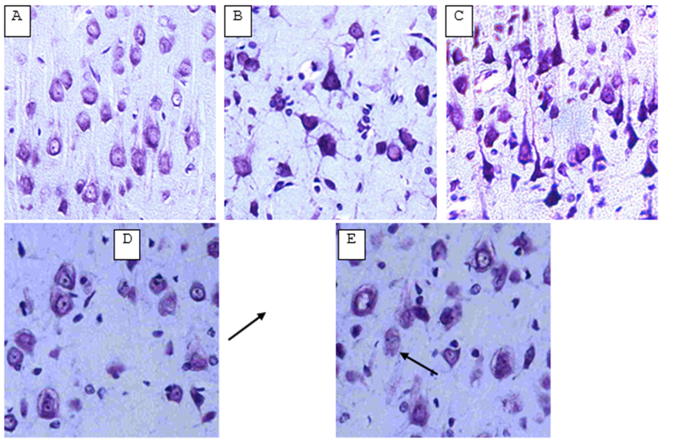
Figure 1: Neurons of the occipital cortex of rats with anoxia and hypoxia of respiratory genesis. Digital micrograph. Nissl staining. Magnifying lens x 20.
A – control group (normochromic neurons);
B – after 30 minutes of anoxia (hyperchromic shrunken neurons);
C – after 60 minutes of anoxia (hyperchromic shrunken neurons);
D – after 30 minutes of hypoxia (hypochromic neurons with signs of swelling and shadow cells (indicated by arrow));
E – after 60 minutes of hypoxia (hypochromic neurons with signs of swelling and shadow cells (indicated by arrow)).
In contrast to the control group, hyperchromic shrunken neurons predominated in the experimental groups with anoxia in both study periods: up to 75% in the group of rats with 30-minute anoxia (p<0>
with residual tracheal patency of 35% in both studied periods led to an increase in the area of neurons without changing their shape with a simultaneous increase in the number of hypochromic neurons with signs of swelling and shadow cells. These differences are due to different severity of acute oxygen deficiency. In total respiratory hypoxia (anoxia), neuronal changes in the form of hyperchromia with signs of wrinkling are characteristic of coagulation necrosis, while in partial respiratory hypoxia, signs of colliquation necrosis are noted in the form of acute swelling of neurons with total chromatolysis and the formation of shadow cells.
Clearly Auctoresonline and particularly Psychology and Mental Health Care Journal is dedicated to improving health care services for individuals and populations. The editorial boards' ability to efficiently recognize and share the global importance of health literacy with a variety of stakeholders. Auctoresonline publishing platform can be used to facilitate of optimal client-based services and should be added to health care professionals' repertoire of evidence-based health care resources.


