AUCTORES
Globalize your Research

Case Report | DOI: https://doi.org/10.31579/ ijcm.2021/0002
*Corresponding Author: Richmond Ronald Gomes, Associate Professor, Medicine, Ad-din Women’s Medical College Hospital, Dhaka, Bangladesh.
Citation: Richmond R Gomes, A Rare Manifestation of Common Disease: Cardiac Cirrhosis. International Journal of Cardiovascular Medicine. DOI: 10.31579/ ijcm.2021/0002
Copyright: © 2021 Richmond Ronald Gomes, this is an open-access article distributed under the terms of the Creative Commons Attribution License, which permits unrestricted use, distribution, and reproduction in any medium, provided the original author and source are credited.
Received: 21 July 2021 | Accepted: 27 August 2021 | Published: 06 September 2021
Keywords: Congestive hepatopathy, Cardiac Cirrhosis, CLD, COPD, pulmonary hypertension
The relation between diseased heart and liver may manifests as acute liver injury, chronic congestive hepatopathy, even cardiac cirrhosis. Congestive hepatopathy caused from impaired blood return to the right ventricle with increased filling pressure. Chronic liver disease (CLD) is the most frequent presentation of hepatobiliary disease. Very rare cause, like long term right heart failure may also be a cause of underlying disease for CLD. We present a case of a 54-year-old female with cardiac cirrhosis. Initial workup was negative. Later thoracic imaging and echocardiography showed chronic obstructive pulmonary disease (COPD) with evidence of pulmonary hypertension. We will briefly discuss the literature on cardiac causes of liver cirrhosis. Our case will present such a short report or cardio-hepatic relations.
Chronic right sided congestive heart failure may cause chronic liver injury and cirrhosis of liver but is very uncommon. In long term right heart failure there is elevated venous pressure that is transmitted to liver sinusoids via inferior vena cava and hepatic veins. This leads to long term passive congestion and relative ischemia due to poor circulation eventually leading to necrosis and fibrosis of liver predominantly of centrilobular region. Patient generally presents with clinical features of congestive heart failure and portal hypertension but very rarely presents with variceal hemorrhage or encephalopathy [1]. But our case patient presented with evidence of variceal hemorrhage. Also the overall prognosis of cardiac cirrhosis is not well established and treatment of cardiac cirrhosis is mainly aimed at managing underlying heart failure so it becomes important to distinguish it from other cause of cirrhosis1.The timely diagnosis of a cardiac etiology of liver dysfunction is important because such dysfunction is potentially reversible if the underlying cardiac disease is treated before the development of frank cirrhosis [2, 3]
A 54 year old lady, home maker, hailing from rural Bangladesh, not known to have diabetes, hypertension or coronary artery disease, chronic smoker(beedi-local hand-rolled cigarettes) presented with progressively increasing abdominal distension for last 6 months, bilateral leg swelling for 1 month and H/O two episodes of passage of black tarry stool since then. On repeated enquiry she also revealed of chronic cough and breathlessness with winter exacerbation for last 10 years and episodes of pedal edema relieving after local medicine. There was no history of alcohol intake, high risk sexual behavior, jaundice, tuberculosis, long term drug or herbal intake, surgery or blood transfusion. There was no significant family history. On general examination patient was cooperative and well oriented with poor nutrition. Pallor, mild icterus and bi-pedal pitting edema was present. Cyanosis, clubbing, lymphadenopathy were absent. Pulse-70/min regular, normovolumic, normal in character and vessel wall normal. Blood pressure-100/ 70 mmHg. Neck vein was engorged and pulsatile and jugular venous pressure raised. On abdominal examination, abdomen was distended diffusely with eversion of umbilicus and prominent veins in flanks and epigastrium with blood flow from below upwards. Abdominal striae was seen. There were no scar marks. No superficial tenderness present. Splenomegaly was present of 4 c.m., firm, non-tender with smooth surface. Liver was also palpable 2 cm from right costal margin along right mid clavicular line, firm, tender with smooth surface and regular margin. No other lump present. Fluid thrill was present. On cardiovascular examination precordium seemed to be normal. Apex beat in 5th intercostal space 2 cm. Lateral to mid clavicular line normal in character. Thrill or para-sternal heave absent. On auscultation 1st and 2nd heart sound audible with loud pulmonary component of 2nd heart sound. The holosystolic, high-pitched, blowing murmur of tricuspid insufficiency best heard at the lower left sternal border. The murmur intensifies with inspiration and decreases with expiration. On respiratory examination chest bilaterally symmetrical with decreased movement on both sides. Trachea central and no deformity of spine seen. Respiratory rate of 26 /min. With use of accessory muscles seen. Vocal fremitus equal on both side. Hyper-Resonant note heard on percussion. Bilaterally decreased breath sounds with diffuse rhonchi heard over lung fields. Vocal resonance decreased bilaterally. Nervous system examination reveals no abnormality.
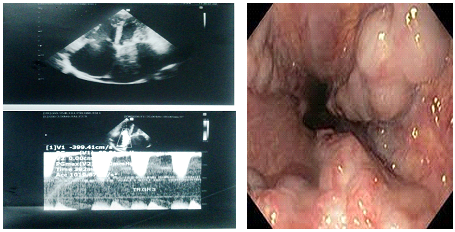
Lab reports revealed: Heamoglobin-9.1 gm/dl,Total leucocyte count-3,800/dl, Differential count-neutrophil-58%, Lymphocytes-30%, Platelets count-117,000/dl. Random Blood sugar-6.6 mmol/L, Serum Sodium-131 mmol/l, Serum Potassium-4.1 mmol/l, Serum Creatinine-0.9 mg/dl Liver Function Test-S. Bilirubin-3.9 gm/dl, SALP-106 IU, SGPT-111 IU, Serum Protien—5.5 gm/dl, Serum Albumin-2.7 gm /dl Ascitic Fluid Examination—TLC-110/ cc, DLC-N35% L65%,Protein-1.3 gm/dl, SAAG-1.4 ADA 12.1U/L(normal lessthan 30U/L) Prothrombin time: patient 18 secs, control 12 secs. Viral markers (HbsAg, HCV)—Negative Chest X-ray-Cardiac Enlargement with accentuation of bronchovascular marking bilateral mild pleural effusion ,ECG-Rate of 70/min with regular rhythm, ABG-pH-7.38 , pCO2-65, pO2-74, SpO2-88% ,USG Abdomen-Liver-17.16 cm, coarse parenchyma, Portal vein-12.9 mm & tortuous, Gross spleenomegaly—15.1 cm., Splenic vein—14.2 mm ,tortuous & dilated with multiple collaterals in perihilar splenic region. Gross peritoneal collection. 2-D Echo-Grade 3 Tricuspid regurgitation, Severe Pulmonary Arterial Hypertension (PASP 40 mm Hg, dilated right ventricle and right atrium (Figure 1). Upper GI Endoscopy-Esophagus shows grade II × III columns of esophageal varices (Figure 2). Pro BNP was 15656 pg/ml (normal lessthan 400 pg/ml). TSH was normal(0.829 IU/ml, normal 0.350-3.40 IU/ml) Pulmonary Function Test-FEV1-52%, FVC-79%, FEV1/FVC-0.66 and improvement in FEV1 after use of bronchodilator was 7% suggesting of chronic obstructive airway disease stage II of GOLD criteria. Fibroscan of liver; Median stiffness was 37.4 Kpa, IQR/MED-10%, which correlate with stage-4 fibrosis, that is cirrhosis. Our final diagnosis was cardiac cirrhosis.
She was started salt and fluid restrictions (daily 1000 ml/day) along with oral diuretics containing frusemide and spironolactone combination. Oral nitrates were advised to prevent further variceal bleeding as b-blockers are avoided in patients with respiratory airway diseases. Long acting b-agonist inhalers, montelukast and doxophyline were given to relieve broncho-constriction. Proton pump inhibitor prescribed to reduce acid production and prevent further damage due to acid reflux. Lactulose prescribed to prevent constipation and related complications. She was also transfused with 1 unit packed red cells and 4 units of fresh frozen plasma. Patient was given education regarding diet, precautions and follow up after discharge.
Term cardiac cirrhosis denotes any type of hepatic fibrosis occurring in cardiac patient [4]. Our case report is in agreement with the previous observations of chronic liver injury due to long term congestive heart failure. It is a very uncommon cause of CLD and it’s difficult to distinguish from other causes of liver cirrhosis. The most important mechanisms responsible for the development of congestive hepatopathy are hepatic congestion, decreased hepatic blood flow and hypoxemia [5] followed by atrophy, necrosis of hepatocytes, thrombi resulting due to cholestasis [6]. Causes of cardiac cirrhosis are valvular heart disease, cardiomyopathy, pericardial disease, ischemic heart disease, primary lung disease [7]. With decrease in incidence valvular heart disease, cardiomyopathy in etiology of cardiac cirrhosis has increased [8].
Our case had primary lung disease due to chronic smoking which resulted in pulmonary hypertension leading to chronic congestive heart failure. This further leads to passive congestion and relative ischemia due to poor circulation eventually leading to necrosis and fibrosis of liver predominantly of centrilobular region [9]. Usually cases of cardiac cirrhosis do not develop variceal hemorrhage or encephalopathy but our case had unusual presentation of melana suggesting variceal bleeding. Our case had Obstructive airway disease of stage II according to GOLD [10] staging evidenced from deranged Pulmonary Function Test, Abnormal Blood Gas analysis. Evidence of Pulmonary hypertension was evident clinically in form of loud P2 and murmur of tricuspid regurgitation which was established on 2D Echocardiography. Chronic congestive heart failure established on long history of 5 years for which he taking treatment (? diuretics) from quack of which records were not available and raised pro-BNP level.
Later he developed congestive hepatopathy and signs of portal hypertensionas evidenced by splenomegaly, progressive ascites which was transudative with SAAG> 1.1 [13], jaundice, dyspnea, engorged neck vein, hepatomegaly, pedal oedema, normal alkaline phosphatase levels, raised AST, ALT and serum bilirubin. Metabolic and synthetic functions of liver were also compromised evident from decreased serum albumin and deranged PT/INR7. In congestive hepatopathy, liver function tests do not show the specific pattern as in patient with hypoxic hepatopathy [11]. Cholestatic enzymes together with low albumin and high bilirubin are the strongest risk factor for poor outcome, in case of chronic heart failure [12]. Chest X-ray was suggestive of congestive cardiac failure as there was bilateral pleural effusion. Splenomegaly was associated with hypersplenism as evident from pancytopenia in blood picture. As our patient was suffering from chronic congestive heart failure and ascites, transabdominal liver biopsy is at risk and transjaugular liver biopsy is not practiced at our setting for the evaluation of cirrhosis. So, fibro scan was done and result was suggesting liver cirrhosis. Usually cases of cardiac cirrhosis does not develop variceal bleeding, but our case presented with variceal bleeding evident from history of melana which was established on upper gastro-intestinal endoscopy in which therapeutic banding could not be done due to financial constraints.
The timely diagnosis of a cardiac etiology of liver dysfunction is important because such dysfunction is potentially reversible if the underlying cardiac disease is treated before the development of frank cirrhosis [2, 3]. Moreover, early treatment of underlying cardiac disease might also prevent the development of hepatocellular carcinoma as suggested by an interesting case study in which a patient with negative hepatitis serologies and cirrhosis secondary to constrictive pericarditis developed hepatocellular carcinoma confirmed by biopsy [14].
This case study illustrates to gastroenterologists the need to consider a cardiac etiology in the work-up of cirrhosis especially when the most common causes are not found .A patient with COPD developing chronic right sided heart failure due to pulmonary hypertension causes passive congestion on hepatic veins, eventually lead to hepatic fibrosis and raised portal hypertension. Though variceal bleed is uncommon in portal hypertension due to cardiac cirrhosis but may be presenting complain in rare case as seen in our case. Thought COPD and cardiac cirrhosis both are very uncommon, our interest was to highlight the cardiac cause should be evaluated in a dysphonic adult, where the causes of CLD were not certain.
None
Clearly Auctoresonline and particularly Psychology and Mental Health Care Journal is dedicated to improving health care services for individuals and populations. The editorial boards' ability to efficiently recognize and share the global importance of health literacy with a variety of stakeholders. Auctoresonline publishing platform can be used to facilitate of optimal client-based services and should be added to health care professionals' repertoire of evidence-based health care resources.


