AUCTORES
Globalize your Research

Case Report | DOI: https://doi.org/10.31579/2640-1053/233
1Department of Pathological Anatomy, Arrazi Hospital, University Hospital of Mohammed VI - Marrakech / Faculty of Medicine and Pharmacy of Marrakech - Cadi Ayyad University Marrakech/Morocco.
2Guelmim Faculty of Medicine and Pharmacy - Ibn Zohr Agadir University Guelmim- Morocco 81000.
3Department of Radiology, Arrazi Hospital, University Hospital of Mohammed VI - Marrakech / Faculty of Medicine and Pharmacy of Marrakech - Cadi Ayyad University Marrakech/Morocco.
4Department of Otorhinolaryngology, University Hospital of Mohammed VI - Marrakech / Faculty of Medicine and Pharmacy of Marrakech - Cadi Ayyad University Marrakech/Morocco.
*Corresponding Author: Imane Boujguenna, Guelmim Faculty of Medicine and Pharmacy - Ibn Zohr Agadir University Guelmim- Morocco.
Citation: Hind Rachidi, Imane Boujguenna, Asma Lahouaoui, Abdeljalil Ouaaziz, Idrissi Guannouni NC, et al, (2025), A Moroccan Chu's Experience with Salivary Gland Tumors, J. Cancer Research and Cellular Therapeutics. 9(2); DOI:10.31579/2640-1053/233
Copyright: © 2025, Imane Boujguenna. This is an open-access article distributed under the terms of the Creative Commons Attribution License, which permits unrestricted use, distribution, and reproduction in any medium, provided the original author and source are credited.
Received: 28 February 2025 | Accepted: 10 March 2025 | Published: 17 March 2025
Keywords: melanoma; choroid; histology; immunohistochemistry
Choroidal melanoma (CM) is malignant neoplasm of melanocytes in choired. It is the most common intraocular malignant neoplasm in adults. The diagnosis is based on histological and immunohistochemical examination. These tumors have a pejorative prognosis and their evolution is marked by the appearance of metastases especially in the liver. Currently, the development of genomic analyzes has led to a better understanding of the molecular mechanisms involved in oncogenesis. Therapeutic management has become much more difficult since the appearance of conservative treatments to keep the eye. We report a new case of choroidal melanoma in a 29-year-old patient diagnosed at the pathology department of the Mohammed VI CHU (Marrakech, Morocco). The aim of this work is to study the anatomo-clinical and evolutionary aspects of this infrequent entity
The salivary gland tumors (SGT) are very different: The OMS 2017 classification recognized 24 subtypes of cancer and 12 subtypes of benign tumors (1). Three 3% of all body tumeurs and 6% of head and cou tumeurs are represented by them. Pléomorphe (AP) is the most common histological type, and parotidienne tumors predominate in their frequency (2,3). Epidemiological data are quite variable, according to studies (4–5).
Establishing an epidemiological profile of salivary gland tumors in a Moroccan environment and comparing it to data from the literature were the goals of this retrospective study.
This is a retrospective study that was carried out over a 16-year period, from January 2008 to September 2024. A total of four venomous patients were taken in charge of a salivary gland tumor. Tumors have been categorized in accordance with OMS 2022 (1). The following factors have been examined: age, sex, location, and histological type of tumors. The EPI INFO 6.0W software was used to collect, code, and analyze all of the data.
There were 42 women and 54 men in the group ; the sex ratio was 1.28, with a little male preponderance. The average age was 42 years old, with extremes ranging from 14 to 83 years old. Forty-five percent of the tumors were parotidiennes, twenty-eight percent were located at the level of the accessory salivary glands, and twenty-three percent were submandibulaires. Eighty-four percent of parotidienne tumors were benign. With an average age of 39 years (14–83), AP accounted for 66.66% of bénigne tumors, followed by cystadénolymphome (CAL) or Warthin tumors, which accounted for 15.55%. The remaining bénigne tumors accounted for 2.22 percent of the cases (fig. 1). The most common malignant form, accounting for 71.42% of cases, was adenoid kystique cancer (CAK), which was followed by mucoépidermoide cancer, which accounted for 40% of cases.
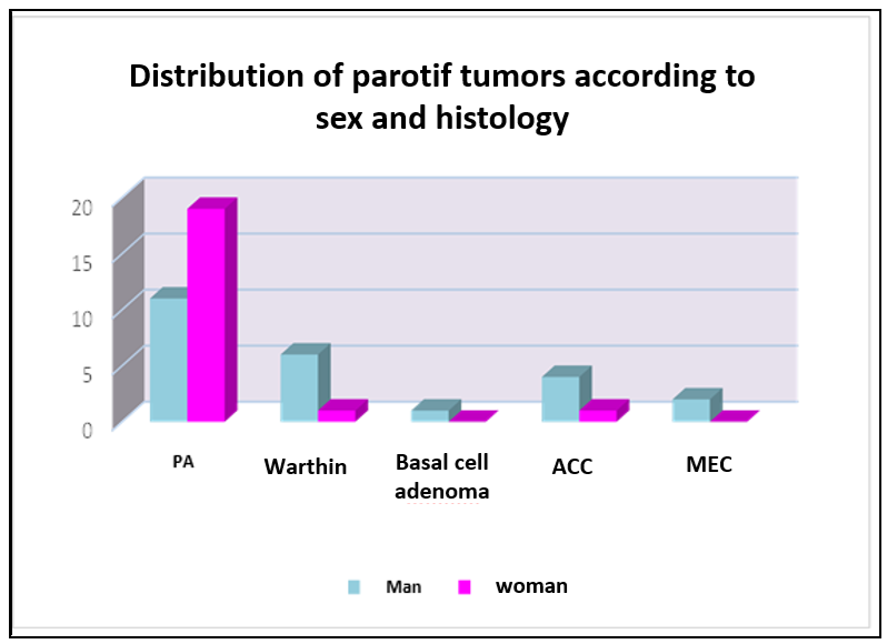
PA: Pleomorphic adenoma - ACC: Adenoid cystic carcinoma - MEC: Mucoepidermoid carcinoma
Figure 1: Distribution of parotid tumors by sex and histological type
Seventy-three percent of submandibulary tumors were bénignes (figure. 2), which were only identified by the term pléomorphe. The CME and CAK represented 66.66% and 33.33% of malignant tumors, respectively.

PA: Pleomorphic adenoma - ACC: Adenoid cystic carcinoma - MEC: Mucoepidermoid carcinoma
Figure 2: Histological type and sex-specific recurrence of tumors under mandibularies
The tumors of the accessory salivary glands (ASG) were all benign and mostly settled at the 70% palatin level. The other locations were the lèvres (8%), the plancher buccal (12%), and the play (10%).
| Nature | Histological Type | Cases number |
Benign tumors (87%) | Pleomorphic adénoma | 75 |
| Cystadénolymphoma | 7 | |
| Basal cell adenoma | 1 | |
Maligned Tumors (13%)
| adenoid cystic carcinoma | 7 |
| mucoépidermoid carcinoma | 6 |
Table 1: Distribution of salivary gland tumors by histological type
The histological diagnosis was performed on biopsy in 19.1%, on surgical specimen in 46.8%, and on excisional biopsy in 34% of cases.
The immunohistochemical study was performed in 11% of the cases. Six cases of recurrence were observed, including three pleomorphic adenomas, one CAK, and one Warthin's tumor.
The average age of occurrence of salivary gland tumors in our context is 42 years, which is consistent with the data in the literature [4,6,8,9].
There is variability in the overall sex ratio according to studies, with either a male predominance as in our study (7), a female predominance [4,6,8], or no sexual predominance [9].
In our study, the parotid location is the predominant one (46.87%), which corresponds to the data in the literature where Parotid involvement ranges from 50 to 83%, submandibular involvement from 5 to 25%, and ASG involvement from 3.2 to 33% [5,6,8,9].
In our series as well as in the majority of studies, salivary gland tumors are most often benign and rarely malignant (Table 1). Malignant tumors occur at an older age [9], and affect men more than women [6,7]. Which is consistent with the results of our study.
Pleomorphic adenoma is the most common benign tumor in our study as well as in other studies in the literature. it most often occurs in women and at a young age (3,5,6,10) Cystadenolymphoma is almost exclusively parotid and represents 5 to 12% of benign salivary gland tumors [10]. In our study, its percentage has slightly increased, reaching 16.66% of our patients.
MEC and ACC are the most common malignant tumors of the salivary glands. CME represents approximately 16% of salivary gland tumors, 44% of malignant tumors [10]. In our study, we note a lower percentage of MEC (4.16%). The CAK represents 5 to 10% of salivary gland tumors, which is consistent with our results.
Salivary gland tumors represent a heterogeneous group of diseases with complex characterization and variable frequency.

Figure 1: (HE*20): benign tumor proliferation of the parotid gland arranged in tubes, often cystic, suggesting a Warthin tumor
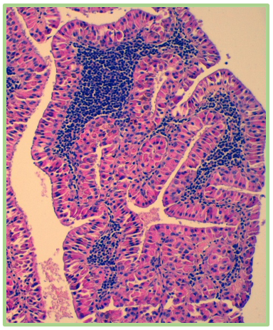
Figure 2: (HE*40): benign tumor proliferation of the parotid gland arranged in tubes, often cystic, suggesting a Warthin's tumor

Figure 3: (HE*20) High-grade infiltrating adenoid cystic carcinoma of the parotid gland in a 70-year-old patient.

Figure 4: (HE*40) High-grade infiltrating adenoid cystic carcinoma of the parotid gland in a 70-year-old patient.

Figure 5: HE*4 infiltrating mucoepidermoid carcinoma affecting the parotid gland.

Figure 6: HE*40 infiltrating mucoepidermoid carcinoma affecting the parotid gland.

Figure 7: HE*4 benign tumor proliferation suggesting a pleomorphic adenoma of the parotid gland.

Figure 8: HE*20 pleomorphic adenoma of the parotid gland.
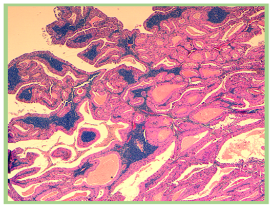
Figure 1: (HE*20): benign tumor proliferation of the parotid gland arranged in tubes, often cystic, suggesting a Warthin tumor

Figure 2: (HE*40): benign tumor proliferation of the parotid gland arranged in tubes, often cystic, suggesting a Warthin's tumor
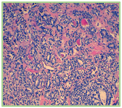
Figure 3: (HE*20) High-grade infiltrating adenoid cystic carcinoma of the parotid gland in a 70-year-old patient.
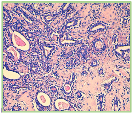
Figure 4: (HE*40) High-grade infiltrating adenoid cystic carcinoma of the parotid gland in a 70-year-old patient.
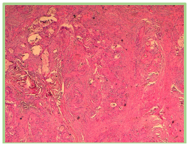
Figure 5: HE*4 infiltrating mucoepidermoid carcinoma affecting the parotid gland.
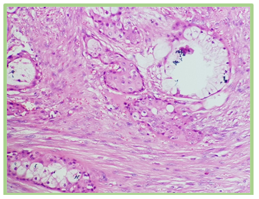
Figure 6: HE*40 infiltrating mucoepidermoid carcinoma affecting the parotid gland.
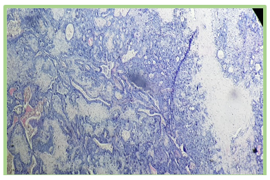
Figure 7: HE*4 benign tumor proliferation suggesting a pleomorphic adenoma of the parotid gland.
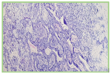
Figure 8: HE*20 pleomorphic adenoma of the parotid gland.
The authors declare that they have no conflicts of interest.
All authors contributed to the writing of this manuscript.
Written informed consent has been obtained from the patient for the publication of this case report and all accompanying images. A copy of the written consent is available for review by the editor-in-chief of this journal.
The authors did not receive any specific funding for this study.
Clearly Auctoresonline and particularly Psychology and Mental Health Care Journal is dedicated to improving health care services for individuals and populations. The editorial boards' ability to efficiently recognize and share the global importance of health literacy with a variety of stakeholders. Auctoresonline publishing platform can be used to facilitate of optimal client-based services and should be added to health care professionals' repertoire of evidence-based health care resources.


