AUCTORES
Globalize your Research
Research Article | DOI: https://doi.org/http://dx.doi.org/10.31579/2019/016
*Corresponding Author: Abdullaiev R.Ya, Kharkov Medical Academy of Postgraduate Education, Ukraine
Citation: Abdullaiev R.Ya. , Kulikova F.I., Alla G. Kyrychenko,Baibakov V., and Dihtiar V. A. Ultrasonic Imaging of Lumbar Degenerative Disc Disease, Journal of Spinal Diseases and Research . DOI: http://dx.doi.org/ 10.31579/ 2019 /016
Copyright: © 2019 Abdullaiev R.Ya This is an open-access article distributed under the terms of the Creative Commons Attribution License, which permits unrestricted use, distribution, and reproduction in any medium, provided the original author and source are credited.
Received: 14 February 2019 | Accepted: 14 March 2019 | Published: 20 March 2019
Keywords: radiculopathy,myelopathy, degenerative disc disease.
The vast majority of radiculopathy and myelopathy in the lumbar spine occurs as a result of spondylosis and degenerative disc disease [1]. It has been established that genetic factors, inflammation, increased body weight play an important role in the development of degenerative disc disease [2,3]. Large-scale studies conducted by a large group of authors - Armbrecht G., Felsenberg D., Ganswindt M. et al. (2017) revealed a close relationship between degenerative disc disease and osteoporosis [4].
The vast majority of radiculopathy and myelopathy in the lumbar spine occurs as a result of spondylosis and degenerative disc disease [1]. It has been established that genetic factors, inflammation, increased body weight play an important role in the development of degenerative disc disease [2,3]. Large-scale studies conducted by a large group of authors - Armbrecht G., Felsenberg D., Ganswindt M. et al. (2017) revealed a close relationship between degenerative disc disease and osteoporosis [4]. Lumbar degenerative disc disease is a multifactorial process, characterized by changes in the architecture and integrity of the disc, which can lead to dysfunction of the involved vertebral motor segment. Dysfunction caused by impaired oxygenation, nutrition, microvasculature and/or inflammation, contribute to the dehydration of the disc, reducing the height of the disc and breaks the annulus fibrous [5]. These processes can improve the innervation of the inner ring and nucleus pulpous with increased stimulation of the dorsal root segmental ganglia (via sinuvertebral nerves) and the paravertebral sympathetic fibers [6]. In studies of Muraki S, Oka H, Akune T et. et al. (2009) showed that narrowing of the disk space and the presence of osteophytes in the intervertebral joints is one of causes lower back pain in spondylosis [7]. Due to central or foraminal stenosis, chronic low back pain may be accompanied by radicular symptoms. Confirmation of the clinical diagnosis of discogenic low back pain using imaging techniques is crucial for deciding the feasibility of surgical treatment of lumbar degenerative disc disease [8].
Studies conducted by Sasiadek MJ and Bladowska J. (2012) showed a high sensitivity of magnetic resonance imaging in the diagnosis of degenerative changes in the intervertebral discs [9]. At the same time, the results of magnetic resonance imaging do not always confirm the direct connection between the intensity of back pain and the degree of degenerative changes in the intervertebral disks. For example, Berg et al. (2013) did not reveal a high correlation between tomographic symptoms of severe degenerative changes in 170 discs and the intensity of pain in patients who were a candidates for prosthesis discs [10]. Takatalo et al. (2012) showed a high correlation between disc herniation and intense back pain. The same authors found that in every third case degenerative changes can be detected in asymptomatic young patients(under 21) who were examined for another reason [11]. Steffens D, et al. (2014) analyzed the results of 12 studies on MRI in the diagnosis of degenerative disc disease and did not reveal a close relationship between it and chronic low back pain [12]. The study of literature data on the diagnosis of degenerative diseases of the disc using imaging methods allowed to make Brinjikji, W. et al. (2015) conclusion: changes in the intervertebral discs detected during an MRI can be age-related, therefore interpretation of the data should be based on clinical symptoms [13].
To study the possibilities of ultrasonic imaging in degenerative lumbar disc disease.
Materials and Methods
A retrospective analysis of ultrasound of the lumbar intervertebral discs was carried out in 49 patients with protrusion and 34 patients with herniated discs at the age of 19-58 years diagnosed by magnetic resonance imaging. The age range of the patients was 19 to 58 years. In all patients were observed clinical symptoms of low back pain of different type: lumbago, radicular pain, radiculopathy as a only sciatica or sciatica in combination with inflammatory changes in epidural space. At the level of 21 discs, spinal canal stenosis was diagnosed. The comparative group consisted of 36 patients without chronic back pain.
The ultrasonic examination was carried out on Philips HD-11 scanners by convexr transducers with a frequency of 2-5 MHz by means of polyprojection and polyposive scanning. The following ultrasonographic symptoms were assessed: homogeneity, echogenicity, integrity of nucleus pulpous, the presence of a protrusion of a disk with or without a fibrous ring rupture, the sagittal size of the spinal canal, the width of the canal of the spinal nerves.
In 19 (38.8%) cases, the protrusion was median, in 23 (46.9%) - paramedian and in 7 (14.3%) – posterolateral. In the group of patients with disc hernia, these parameters were: 18 (52.9), 12 (35,3%) and 4 (11,8%), respectively.
Low back pain was characterized as a Lumbago and Sciatica. Lumbago among patients with disc protrusion was observed in 31 (63,2%) cases – from them in 24 (49,0%) cases with median, in 5 (10,2%) cases paramedian and in 1 (2,0%) cases with posterolateral localization. Sciatica was observed in 18 (36,8%) cases – from them in 2 (4,1%) cases with median, in 5 (10,2%) cases paramedian and in 11 (22,4%) cases with posterolateral localization.
Lumbago among patients with disc hernia was observed in 12 (35,3%) cases – from them in 4 (11,8%) cases with median, in 3 (8,8%) cases paramedian and in 5 (14,7%) cases with posterolateral localization. Sciatica was observed in 22 (64,7%) cases – from them in 2 (5,9%) cases with median, in 7 (20,6%) cases paramedian and in 13 (38,2%) cases with posterolateral localization. The lumbago was significantly more frequent in patients with median protrusions, but sciatica – on the contrary, in posterolateral localization of hernia (P < 0,01).
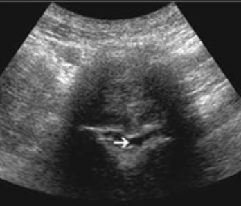
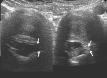
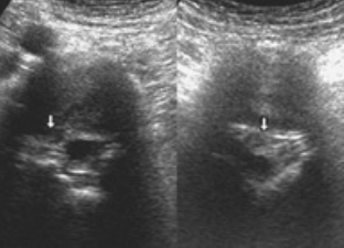
Degenerative changes in the discs, especially the formation of a hernia, are the most frequent causes of spinal pain. The postlateral hernias and protrusions cause a maximal decrease in the width of the spinal nerve canal and compression of nerve root (Figure 1-3).


At the L2-L3 level, the root canals are symmetrical (left arrows), at the level L1-L2, a left-side paramedical protrusion with a stenosis of the root canal (right arrows).

1. Ultrasonography is an effective method for diagnosing degenerative-dystrophic changes in the motor segment of a vertebra and can help in determining the pathophysiological mechanism of back pain.
2. The availability, low cost of ultrasonographic examination allows using the method not only as a screening for degenerative changes in lumbar intervertebral discs, but also for differentiating the causes of back pain in combination with clinical symptoms.
3. The smallest width of the spinal nerve canal is observed with posterolateral hernias and Sciatica is more often observed in these patients.